 Movie
Movie Controller

[English] 日本語
 Yorodumi
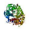
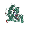
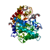
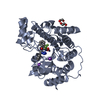
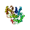
- PDB-6sbf: Structure of type II terpene cyclase MstE_Y157F from Scytonema (apo) -
+ Open data

- Basic information
| Entry | Database: PDB / ID: 6sbf | |||||||||
|---|---|---|---|---|---|---|---|---|---|---|
| Title | Structure of type II terpene cyclase MstE_Y157F from Scytonema (apo) | |||||||||
 Components Components | MstE | |||||||||
 Keywords Keywords | BIOSYNTHETIC PROTEIN / Type II Terpene Cyclase / Marine Drugs / Merosterol / Alpha6-Alpha6 Barrel | |||||||||
| Function / homology | Squalene cyclase, C-terminal / Squalene-hopene cyclase C-terminal domain / Terpenoid cyclases/protein prenyltransferase alpha-alpha toroid / BETA-MERCAPTOETHANOL / MstE Function and homology information Function and homology information | |||||||||
| Biological species |  Scytonema sp. PCC 10023 (bacteria) Scytonema sp. PCC 10023 (bacteria) | |||||||||
| Method |  X-RAY DIFFRACTION / SYNCHROTRON / MOLECULAR REPLACEMENT / Resolution: 1.3 Å X-RAY DIFFRACTION / SYNCHROTRON / MOLECULAR REPLACEMENT / Resolution: 1.3 Å | |||||||||
 Authors Authors | Moosmann, P. / Ecker, F. / Leopold-Messer, S. / Cahn, J.K.B. / Groll, M. / Piel, J. | |||||||||
| Funding support |  Switzerland, 2items Switzerland, 2items
| |||||||||
 Citation Citation | Journal: Nat.Chem. / Year: 2020 Title: A monodomain class II terpene cyclase assembles complex isoprenoid scaffolds. Authors: Moosmann, P. / Ecker, F. / Leopold-Messer, S. / Cahn, J.K.B. / Dieterich, C.L. / Groll, M. / Piel, J. | |||||||||
| History |
|
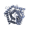
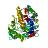
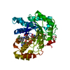
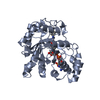
- Structure visualization
| Structure viewer | Molecule: MolmilJmol/JSmol |
|---|
- Downloads & links
-Download
| PDBx/mmCIF format | 6sbf.cif.gz | 165.4 KB | Display | PDBx/mmCIF format |
|---|---|---|---|---|
| PDB format | pdb6sbf.ent.gz | 129.2 KB | Display | PDB format |
| PDBx/mmJSON format | 6sbf.json.gz | Tree view | PDBx/mmJSON format | |
| Others |  Other downloads Other downloads |
-Validation report
| Arichive directory | https://data.pdbj.org/pub/pdb/validation_reports/sb/6sbfftp://data.pdbj.org/pub/pdb/validation_reports/sb/6sbf | HTTPS FTP |
|---|
-Related structure data
| Related structure data | 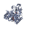 6sbbSC 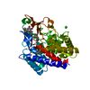 6sbcC 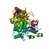 6sbdC  6sbeC  6sbgC S: Starting model for refinement C: citing same article ( |
|---|---|
| Similar structure data |

-Links
 PDBj
PDBj


- Assembly
| Deposited unit | 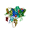
| ||||||||
|---|---|---|---|---|---|---|---|---|---|
| 1 |
| ||||||||
| Unit cell |
|
-Components
| #1: Protein | Mass: 40636.402 Da / Num. of mol.: 1 Source method: isolated from a genetically manipulated source Source: (gene. exp.) Scytonema sp. PCC 10023 (bacteria) / Gene: mstE / Production host: | ||||||
|---|---|---|---|---|---|---|---|
| #2: Chemical |   Mass: 92.094 Da / Num. of mol.: 2 / Source method: obtained synthetically / Formula: C3H8O3 Mass: 92.094 Da / Num. of mol.: 2 / Source method: obtained synthetically / Formula: C3H8O3#3: Chemical | ChemComp-BME / |   Mass: 78.133 Da / Num. of mol.: 1 / Source method: obtained synthetically / Formula: C2H6OS Mass: 78.133 Da / Num. of mol.: 1 / Source method: obtained synthetically / Formula: C2H6OS#4: Water | ChemComp-HOH / |  Mass: 18.015 Da / Num. of mol.: 326 / Source method: isolated from a natural source / Formula: H2O Mass: 18.015 Da / Num. of mol.: 326 / Source method: isolated from a natural source / Formula: H2OHas ligand of interest | N | |
-Experimental details
-Experiment
| Experiment | Method: X-RAY DIFFRACTION / Number of used crystals: 1 |
|---|
- Sample preparation
| Crystal | Density Matthews: 2.08 Å3/Da / Density % sol: 40.88 % |
|---|---|
| Crystal grow | Temperature: 293 K / Method: vapor diffusion, sitting drop / pH: 6.5 / Details: 3.4 M Na-formate |
-Data collection
| Diffraction | Mean temperature: 100 K / Serial crystal experiment: N |
|---|---|
| Diffraction source | Source: SYNCHROTRON / Site: SLS / Beamline: X06SA / Wavelength: 1 Å |
| Detector | Type: DECTRIS EIGER X 16M / Detector: PIXEL / Date: Dec 1, 2018 |
| Radiation | Protocol: SINGLE WAVELENGTH / Monochromatic (M) / Laue (L): M / Scattering type: x-ray |
| Radiation wavelength | Wavelength: 1 Å / Relative weight: 1 |
| Reflection | Resolution: 1.3→50 Å / Num. obs: 83943 / % possible obs: 99.8 % / Redundancy: 5.1 % / Rmerge(I) obs: 0.044 / Net I/σ(I): 16.69 |
| Reflection shell | Resolution: 1.3→1.4 Å / Rmerge(I) obs: 0.562 / Mean I/σ(I) obs: 2.7 / Num. unique obs: 16531 / % possible all: 99.9 |
- Processing
| Software |
| |||||||||||||||||||||||||||||||||||||||||||||||||||||||||||||||||
|---|---|---|---|---|---|---|---|---|---|---|---|---|---|---|---|---|---|---|---|---|---|---|---|---|---|---|---|---|---|---|---|---|---|---|---|---|---|---|---|---|---|---|---|---|---|---|---|---|---|---|---|---|---|---|---|---|---|---|---|---|---|---|---|---|---|---|
| Refinement | Method to determine structure: MOLECULAR REPLACEMENT Starting model: 6SBB Resolution: 1.3→15 Å / Cor.coef. Fo:Fc: 0.985 / Cor.coef. Fo:Fc free: 0.98 / SU B: 1.669 / SU ML: 0.03 / Cross valid method: THROUGHOUT / σ(F): 0 / ESU R: 0.037 / ESU R Free: 0.039 Details: HYDROGENS HAVE BEEN ADDED IN THE RIDING POSITIONS U VALUES : REFINED INDIVIDUALLY
| |||||||||||||||||||||||||||||||||||||||||||||||||||||||||||||||||
| Solvent computation | Ion probe radii: 0.8 Å / Shrinkage radii: 0.8 Å / VDW probe radii: 1.2 Å | |||||||||||||||||||||||||||||||||||||||||||||||||||||||||||||||||
| Displacement parameters | Biso max: 114.35 Å2 / Biso mean: 19.27 Å2 / Biso min: 10.36 Å2
| |||||||||||||||||||||||||||||||||||||||||||||||||||||||||||||||||
| Refinement step | Cycle: final / Resolution: 1.3→15 Å
| |||||||||||||||||||||||||||||||||||||||||||||||||||||||||||||||||
| Refine LS restraints |
| |||||||||||||||||||||||||||||||||||||||||||||||||||||||||||||||||
| LS refinement shell | Resolution: 1.3→1.334 Å / Rfactor Rfree error: 0 / Total num. of bins used: 20
|
