 Movie
Movie Controller

[English] 日本語
 Yorodumi
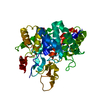
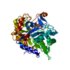
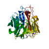
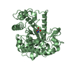
- PDB-2whm: Cellvibrio japonicus Man26A E121A and E320G double mutant in comp... -
+ Open data

- Basic information
| Entry | Database: PDB / ID: 2whm | |||||||||
|---|---|---|---|---|---|---|---|---|---|---|
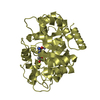
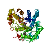
| Title | Cellvibrio japonicus Man26A E121A and E320G double mutant in complex with mannobiose | |||||||||
 Components Components | ENDO-1,4-BETA MANNANASE, MAN26A | |||||||||
 Keywords Keywords | HYDROLASE / GLYCOSIDE HYDROLASE / MAN26 / GH-A CLAN / MANNANASE / GLYCOSIDASE | |||||||||
| Function / homology |  Function and homology information Function and homology informationglucomannan metabolic process / galactomannan metabolic process / mannan endo-1,4-beta-mannosidase / mannan endo-1,4-beta-mannosidase activity / polysaccharide catabolic process Similarity search - Function | |||||||||
| Biological species |  CELLVIBRIO JAPONICUS (bacteria) CELLVIBRIO JAPONICUS (bacteria) | |||||||||
| Method |  X-RAY DIFFRACTION / SYNCHROTRON / MOLECULAR REPLACEMENT / Resolution: 1.5 Å X-RAY DIFFRACTION / SYNCHROTRON / MOLECULAR REPLACEMENT / Resolution: 1.5 Å | |||||||||
 Authors Authors | Durcos, V.M.A. / Davies, G.J. / Flint, J.E. / Gilbert, H.J. | |||||||||
 Citation Citation | Journal: Biochemistry / Year: 2009 Title: Understanding How Diverse -Mannanases Recognise Heterogeneous Substrates. Authors: Tailford, L.E. / Ducros, V.M.A. / Flint, J.E. / Roberts, S.M. / Morland, C. / Zechel, D.L. / Smith, N. / Bjornvad, M.E. / Borchert, T.V. / Wilson, K.S. / Davies, G.J. / Gilbert, H.J. | |||||||||
| History |
| |||||||||
| Remark 700 | SHEET THE SHEET STRUCTURE OF THIS MOLECULE IS BIFURCATED. IN ORDER TO REPRESENT THIS FEATURE IN ... SHEET THE SHEET STRUCTURE OF THIS MOLECULE IS BIFURCATED. IN ORDER TO REPRESENT THIS FEATURE IN THE SHEET RECORDS BELOW, TWO SHEETS ARE DEFINED. |
- Structure visualization
| Structure viewer | Molecule: MolmilJmol/JSmol |
|---|
- Downloads & links
-Download
| PDBx/mmCIF format | 2whm.cif.gz | 181.2 KB | Display | PDBx/mmCIF format |
|---|---|---|---|---|
| PDB format | pdb2whm.ent.gz | 141.4 KB | Display | PDB format |
| PDBx/mmJSON format | 2whm.json.gz | Tree view | PDBx/mmJSON format | |
| Others |  Other downloads Other downloads |
-Validation report
| Arichive directory | https://data.pdbj.org/pub/pdb/validation_reports/wh/2whmftp://data.pdbj.org/pub/pdb/validation_reports/wh/2whm | HTTPS FTP |
|---|
-Related structure data
| Related structure data | 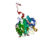 2whjC 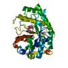 2whkC 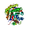 2whlC 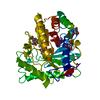 1gw1S C: citing same article ( S: Starting model for refinement |
|---|---|
| Similar structure data |


-Links
 PDBj
PDBj- Assembly
| Deposited unit | 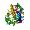
| ||||||||
|---|---|---|---|---|---|---|---|---|---|
| 1 |
| ||||||||
| Unit cell |
|
-Components
-Protein / Sugars , 2 types, 2 molecules A
| #1: Protein | Mass: 43486.352 Da / Num. of mol.: 1 / Fragment: RESIDUES 39-423 / Mutation: YES Source method: isolated from a genetically manipulated source Source: (gene. exp.) CELLVIBRIO JAPONICUS (bacteria) / Production host: References: UniProt: B3PBK3, UniProt: P49424*PLUS, mannan endo-1,4-beta-mannosidase |
|---|---|
| #2: Polysaccharide | beta-D-mannopyranose-(1-4)-alpha-D-mannopyranose Source method: isolated from a genetically manipulated source |
-Non-polymers , 4 types, 427 molecules 


| #3: Chemical | ChemComp-ZN /  Mass: 65.409 Da / Num. of mol.: 1 / Source method: obtained synthetically / Formula: Zn Mass: 65.409 Da / Num. of mol.: 1 / Source method: obtained synthetically / Formula: Zn | ||
|---|---|---|---|
| #4: Chemical | ChemComp-NA /  Mass: 22.990 Da / Num. of mol.: 1 / Source method: obtained synthetically / Formula: Na Mass: 22.990 Da / Num. of mol.: 1 / Source method: obtained synthetically / Formula: Na | ||
| #5: Chemical |  Mass: 122.143 Da / Num. of mol.: 2 / Source method: obtained synthetically / Formula: C4H12NO3 / Comment: pH buffer*YM Mass: 122.143 Da / Num. of mol.: 2 / Source method: obtained synthetically / Formula: C4H12NO3 / Comment: pH buffer*YM#6: Water | ChemComp-HOH / | Mass: 18.015 Da / Num. of mol.: 423 / Source method: isolated from a natural source / Formula: H2O |
-Details
| Nonpolymer details | MANNOSE (MAN): FOUND AS PART OF A BETA 1-4 LINKED MNNOBIOSID| Sequence details | ENZYME IS DOUBLE VARIANT E121A E320G | |
|---|
-Experimental details
-Experiment
| Experiment | Method: X-RAY DIFFRACTION / Number of used crystals: 1 |
|---|
- Sample preparation
| Crystal | Density Matthews: 2.7 Å3/Da / Density % sol: 55 % / Description: NONE |
|---|---|
| Crystal grow | pH: 7.5 Details: 100MMTRIS PH7, 26% MEONOMETHYLETHER PEG550, 3MM ZNSO4, pH 7.5 |
-Data collection
| Diffraction | Mean temperature: 100 K |
|---|---|
| Diffraction source | Source: SYNCHROTRON / Site: ESRF  / Beamline: ID14-1 / Wavelength: 0.934 / Beamline: ID14-1 / Wavelength: 0.934 |
| Detector | Type: ADSC CCD / Detector: CCD |
| Radiation | Protocol: SINGLE WAVELENGTH / Monochromatic (M) / Laue (L): M / Scattering type: x-ray |
| Radiation wavelength | Wavelength: 0.934 Å / Relative weight: 1 |
| Reflection | Resolution: 1.5→30 Å / Num. obs: 73813 / % possible obs: 100 % / Observed criterion σ(I): 0 / Redundancy: 3.7 % / Rmerge(I) obs: 0.04 / Net I/σ(I): 53 |
| Reflection shell | Resolution: 1.5→1.55 Å / Redundancy: 3.7 % / Rmerge(I) obs: 0.27 / Mean I/σ(I) obs: 5.2 / % possible all: 100 |
- Processing
| Software |
| ||||||||||||||||||||||||||||||||||||||||||||||||||||||||||||||||||||||||||||||||||||||||||||||||||||||||||||||||||||||||||||||||||||||||||||||||||||||||||||||||||||||||||||||||||||||
|---|---|---|---|---|---|---|---|---|---|---|---|---|---|---|---|---|---|---|---|---|---|---|---|---|---|---|---|---|---|---|---|---|---|---|---|---|---|---|---|---|---|---|---|---|---|---|---|---|---|---|---|---|---|---|---|---|---|---|---|---|---|---|---|---|---|---|---|---|---|---|---|---|---|---|---|---|---|---|---|---|---|---|---|---|---|---|---|---|---|---|---|---|---|---|---|---|---|---|---|---|---|---|---|---|---|---|---|---|---|---|---|---|---|---|---|---|---|---|---|---|---|---|---|---|---|---|---|---|---|---|---|---|---|---|---|---|---|---|---|---|---|---|---|---|---|---|---|---|---|---|---|---|---|---|---|---|---|---|---|---|---|---|---|---|---|---|---|---|---|---|---|---|---|---|---|---|---|---|---|---|---|---|---|
| Refinement | Method to determine structure: MOLECULAR REPLACEMENT Starting model: PDB ENTRY 1GW1 Resolution: 1.5→19.98 Å / Cor.coef. Fo:Fc: 0.974 / Cor.coef. Fo:Fc free: 0.969 / SU B: 1.771 / SU ML: 0.03 / Cross valid method: THROUGHOUT / ESU R: 0.066 / ESU R Free: 0.056 / Stereochemistry target values: MAXIMUM LIKELIHOOD Details: HYDROGENS HAVE BEEN ADDED IN THE RIDING POSITIONS. U VALUES REFINED INDIVIDUALLY
| ||||||||||||||||||||||||||||||||||||||||||||||||||||||||||||||||||||||||||||||||||||||||||||||||||||||||||||||||||||||||||||||||||||||||||||||||||||||||||||||||||||||||||||||||||||||
| Solvent computation | Ion probe radii: 0.8 Å / Shrinkage radii: 0.8 Å / VDW probe radii: 1.4 Å / Solvent model: MASK | ||||||||||||||||||||||||||||||||||||||||||||||||||||||||||||||||||||||||||||||||||||||||||||||||||||||||||||||||||||||||||||||||||||||||||||||||||||||||||||||||||||||||||||||||||||||
| Displacement parameters | Biso mean: 18.25 Å2
| ||||||||||||||||||||||||||||||||||||||||||||||||||||||||||||||||||||||||||||||||||||||||||||||||||||||||||||||||||||||||||||||||||||||||||||||||||||||||||||||||||||||||||||||||||||||
| Refinement step | Cycle: LAST / Resolution: 1.5→19.98 Å
| ||||||||||||||||||||||||||||||||||||||||||||||||||||||||||||||||||||||||||||||||||||||||||||||||||||||||||||||||||||||||||||||||||||||||||||||||||||||||||||||||||||||||||||||||||||||
| Refine LS restraints |
|