 Movie
Movie Controller

[English] 日本語
 Yorodumi
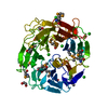
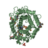
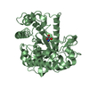
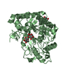
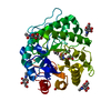
- PDB-1gw1: Substrate distortion by beta-mannanase from Pseudomonas cellulosa -
+ Open data

- Basic information
| Entry | Database: PDB / ID: 1gw1 | |||||||||
|---|---|---|---|---|---|---|---|---|---|---|
| Title | Substrate distortion by beta-mannanase from Pseudomonas cellulosa | |||||||||
 Components Components | MANNAN ENDO-1,4-BETA-MANNOSIDASE | |||||||||
 Keywords Keywords | HYDROLASE / GLYCOSIDE HYDROLASE / GLYCOSIDASE | |||||||||
| Function / homology |  Function and homology information Function and homology informationglucomannan metabolic process / galactomannan metabolic process / mannan endo-1,4-beta-mannosidase / mannan endo-1,4-beta-mannosidase activity / polysaccharide catabolic process Similarity search - Function | |||||||||
| Biological species |  PSEUDOMONAS CELLULOSA (bacteria) PSEUDOMONAS CELLULOSA (bacteria) | |||||||||
| Method |  X-RAY DIFFRACTION / SYNCHROTRON / MOLECULAR REPLACEMENT / Resolution: 1.65 Å X-RAY DIFFRACTION / SYNCHROTRON / MOLECULAR REPLACEMENT / Resolution: 1.65 Å | |||||||||
 Authors Authors | Ducros, V. / Zechel, D.L. / Gilbert, H.J. / Szabo, L. / Withers, S.G. / Davies, G.J. | |||||||||
 Citation Citation | Journal: Angew.Chem.Int.Ed.Engl. / Year: 2002 Title: Substrate Distortion by a Beta-Mannanase: Snapshots of the Michaelis and Covalent-Intermediate Complexes Suggest a B2,5 Conformation for the Transition State Authors: Ducros, V. / Zechel, D.L. / Murshudov, G. / Gilbert, H.J. / Szabo, L. / Stoll, D. / Withers, S.G. / Davies, G.J. #1: Journal: J.Biol.Chem. / Year: 2001Title: Crystal Structure of Mannanase 26A from Pseudomonas Cellulosa and Analysis of Residues Involved in Substrate Binding Authors: Hogg, D. / Woo, E.-J. / Bolam, D.N. / Mckie, V.A. / Gilbert, H.J. / Pickersgill, R.W. | |||||||||
| History |
| |||||||||
| Remark 700 | SHEET THE SHEET STRUCTURE OF THIS MOLECULE IS BIFURCATED. IN ORDER TO REPRESENT THIS FEATURE IN ... SHEET THE SHEET STRUCTURE OF THIS MOLECULE IS BIFURCATED. IN ORDER TO REPRESENT THIS FEATURE IN THE SHEET RECORDS BELOW, TWO SHEETS ARE DEFINED. |
- Structure visualization
| Structure viewer | Molecule: MolmilJmol/JSmol |
|---|
- Downloads & links
-Download
| PDBx/mmCIF format | 1gw1.cif.gz | 182.2 KB | Display | PDBx/mmCIF format |
|---|---|---|---|---|
| PDB format | pdb1gw1.ent.gz | 142.7 KB | Display | PDB format |
| PDBx/mmJSON format | 1gw1.json.gz | Tree view | PDBx/mmJSON format | |
| Others |  Other downloads Other downloads |
-Validation report
| Arichive directory | https://data.pdbj.org/pub/pdb/validation_reports/gw/1gw1ftp://data.pdbj.org/pub/pdb/validation_reports/gw/1gw1 | HTTPS FTP |
|---|
-Related structure data
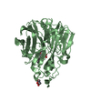
| Related structure data | 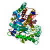 1gvyC  1j9yS C: citing same article ( S: Starting model for refinement |
|---|---|
| Similar structure data |


-Links
 PDBj
PDBj- Assembly
| Deposited unit | 
| ||||||||
|---|---|---|---|---|---|---|---|---|---|
| 1 |
| ||||||||
| Unit cell |
|
-Components
-Protein / Sugars , 2 types, 2 molecules A
| #1: Protein | Mass: 42619.348 Da / Num. of mol.: 1 / Mutation: YES Source method: isolated from a genetically manipulated source Details: COMPLEX WITH 2,4-DINITROPHENYL 2-DEOXY-2-FLUORO-BETA-MANNOTRIOSIDE Source: (gene. exp.) PSEUDOMONAS CELLULOSA (bacteria) / Production host: References: UniProt: P49424, mannan endo-1,4-beta-mannosidase |
|---|---|
| #2: Polysaccharide | beta-D-mannopyranose-(1-4)-beta-D-mannopyranose-(1-4)-2-deoxy-2-fluoro-alpha-D-mannopyranose Source method: isolated from a genetically manipulated source |
-Non-polymers , 5 types, 385 molecules 


| #3: Chemical |  Mass: 65.409 Da / Num. of mol.: 2 / Source method: obtained synthetically / Formula: Zn Mass: 65.409 Da / Num. of mol.: 2 / Source method: obtained synthetically / Formula: Zn#4: Chemical | ChemComp-NA / |  Mass: 22.990 Da / Num. of mol.: 1 / Source method: obtained synthetically / Formula: Na Mass: 22.990 Da / Num. of mol.: 1 / Source method: obtained synthetically / Formula: Na#5: Chemical | ChemComp-NIN / |  Mass: 168.107 Da / Num. of mol.: 1 / Source method: obtained synthetically / Formula: C6H4N2O4 Mass: 168.107 Da / Num. of mol.: 1 / Source method: obtained synthetically / Formula: C6H4N2O4#6: Chemical |  Mass: 122.143 Da / Num. of mol.: 2 / Source method: obtained synthetically / Formula: C4H12NO3 / Comment: pH buffer*YM Mass: 122.143 Da / Num. of mol.: 2 / Source method: obtained synthetically / Formula: C4H12NO3 / Comment: pH buffer*YM#7: Water | ChemComp-HOH / | Mass: 18.015 Da / Num. of mol.: 379 / Source method: isolated from a natural source / Formula: H2O |
|---|
-Details
| Compound details | ENGINEERED| Has protein modification | Y | Sequence details | THE SWISS-PROT ACCESSION NUMBER IS OF THE NATIVE, THE SEQUENCE OF THE STRUCTURE DEPOSITED IS OF A ...THE SWISS-PROT ACCESSION NUMBER IS OF THE NATIVE, THE SEQUENCE OF THE STRUCTURE DEPOSITED IS OF A MUTANT E212A BREAK IN CHAIN A: RESIDUES 370 AND 371 NOT BUILT DUE TO DISORDER IN THE DENSITY | |
|---|
-Experimental details
-Experiment
| Experiment | Method: X-RAY DIFFRACTION / Number of used crystals: 1 |
|---|
- Sample preparation
| Crystal | Density Matthews: 2.67 Å3/Da / Density % sol: 53.62 % | ||||||||||||||||||||
|---|---|---|---|---|---|---|---|---|---|---|---|---|---|---|---|---|---|---|---|---|---|
| Crystal grow | pH: 7.5 / Details: 100MM TRIS 7.5, 9MM ZNSO4, 26% PEG550, pH 7.50 | ||||||||||||||||||||
| Crystal grow | *PLUS pH: 6.5 / Method: vapor diffusion, hanging drop / Details: Hogg, D., (2001) J.Biol.Chem., 276, 31186. | ||||||||||||||||||||
| Components of the solutions | *PLUS
|
-Data collection
| Diffraction | Mean temperature: 100 K |
|---|---|
| Diffraction source | Source: SYNCHROTRON / Site: ESRF  / Beamline: ID14-2 / Wavelength: 0.933 / Beamline: ID14-2 / Wavelength: 0.933 |
| Detector | Type: ADSC CCD / Detector: CCD / Date: Jun 15, 2001 / Details: TOROIDAL MIRROR |
| Radiation | Monochromator: DIAMOND (111), GE(220) / Protocol: SINGLE WAVELENGTH / Monochromatic (M) / Laue (L): M / Scattering type: x-ray |
| Radiation wavelength | Wavelength: 0.933 Å / Relative weight: 1 |
| Reflection | Resolution: 1.65→20 Å / Num. obs: 54188 / % possible obs: 100 % / Redundancy: 4.7 % / Rmerge(I) obs: 0.066 / Net I/σ(I): 19 |
| Reflection shell | Resolution: 1.65→1.71 Å / Redundancy: 4.9 % / Rmerge(I) obs: 0.264 / Mean I/σ(I) obs: 9.3 / % possible all: 99 |
- Processing
| Software |
| ||||||||||||||||||||||||||||||||||||||||||||||||||||||||||||||||||||||||||||||||||||||||||||||||||||||||||||||||||||||||||||||||||||||||||||||||||||||||||||||||||||||||||||||||||||||
|---|---|---|---|---|---|---|---|---|---|---|---|---|---|---|---|---|---|---|---|---|---|---|---|---|---|---|---|---|---|---|---|---|---|---|---|---|---|---|---|---|---|---|---|---|---|---|---|---|---|---|---|---|---|---|---|---|---|---|---|---|---|---|---|---|---|---|---|---|---|---|---|---|---|---|---|---|---|---|---|---|---|---|---|---|---|---|---|---|---|---|---|---|---|---|---|---|---|---|---|---|---|---|---|---|---|---|---|---|---|---|---|---|---|---|---|---|---|---|---|---|---|---|---|---|---|---|---|---|---|---|---|---|---|---|---|---|---|---|---|---|---|---|---|---|---|---|---|---|---|---|---|---|---|---|---|---|---|---|---|---|---|---|---|---|---|---|---|---|---|---|---|---|---|---|---|---|---|---|---|---|---|---|---|
| Refinement | Method to determine structure: MOLECULAR REPLACEMENT Starting model: PDB ENTRY 1J9Y Resolution: 1.65→20 Å / Cor.coef. Fo:Fc: 0.971 / Cor.coef. Fo:Fc free: 0.961 / SU B: 1.263 / SU ML: 0.044 / Cross valid method: THROUGHOUT / ESU R: 0.097 / ESU R Free: 0.073 / Stereochemistry target values: MAXIMUM LIKELIHOOD / Details: HYDROGENS HAVE BEEN ADDED IN THE RIDING POSITIONS
| ||||||||||||||||||||||||||||||||||||||||||||||||||||||||||||||||||||||||||||||||||||||||||||||||||||||||||||||||||||||||||||||||||||||||||||||||||||||||||||||||||||||||||||||||||||||
| Solvent computation | Ion probe radii: 0.8 Å / Shrinkage radii: 0.8 Å / VDW probe radii: 1.4 Å / Solvent model: BABINET MODEL WITH MASK | ||||||||||||||||||||||||||||||||||||||||||||||||||||||||||||||||||||||||||||||||||||||||||||||||||||||||||||||||||||||||||||||||||||||||||||||||||||||||||||||||||||||||||||||||||||||
| Displacement parameters | Biso mean: 18.08 Å2
| ||||||||||||||||||||||||||||||||||||||||||||||||||||||||||||||||||||||||||||||||||||||||||||||||||||||||||||||||||||||||||||||||||||||||||||||||||||||||||||||||||||||||||||||||||||||
| Refinement step | Cycle: LAST / Resolution: 1.65→20 Å
| ||||||||||||||||||||||||||||||||||||||||||||||||||||||||||||||||||||||||||||||||||||||||||||||||||||||||||||||||||||||||||||||||||||||||||||||||||||||||||||||||||||||||||||||||||||||
| Refine LS restraints |
|