 Movie
Movie Controller


+ Open data

- Basic information
| Entry | Database: PDB / ID: 4zvd | ||||||
|---|---|---|---|---|---|---|---|
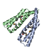
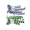
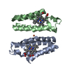
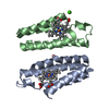
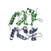
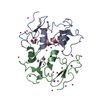
| Title | Crystal structure of MID domain of the E. coli DosC - form II | ||||||
 Components Components | Diguanylate cyclase DosC | ||||||
 Keywords Keywords | SIGNALING PROTEIN / oxygen sensing / diguanylate cyclase / cyclic-di-GMP | ||||||
| Function / homology |  Function and homology information Function and homology informationnegative regulation of bacterial-type flagellum-dependent cell motility / diguanylate cyclase / diguanylate cyclase activity / carbon monoxide binding / response to oxygen levels / cell adhesion involved in single-species biofilm formation / response to stress / oxygen binding / heme binding / GTP binding ...negative regulation of bacterial-type flagellum-dependent cell motility / diguanylate cyclase / diguanylate cyclase activity / carbon monoxide binding / response to oxygen levels / cell adhesion involved in single-species biofilm formation / response to stress / oxygen binding / heme binding / GTP binding / protein homodimerization activity / metal ion binding / plasma membrane Similarity search - Function | ||||||
| Biological species |  | ||||||
| Method |  X-RAY DIFFRACTION / SYNCHROTRON / MOLECULAR REPLACEMENT / Resolution: 1.9 Å X-RAY DIFFRACTION / SYNCHROTRON / MOLECULAR REPLACEMENT / Resolution: 1.9 Å | ||||||
 Authors Authors | Tarnawski, M. / Barends, T.R.M. / Schlichting, I. | ||||||
 Citation Citation | Journal: Acta Crystallogr.,Sect.D / Year: 2015 Title: Structural analysis of an oxygen-regulated diguanylate cyclase. Authors: Tarnawski, M. / Barends, T.R. / Schlichting, I. | ||||||
| History |
|
- Structure visualization
| Structure viewer | Molecule: MolmilJmol/JSmol |
|---|
- Downloads & links
-Download
| PDBx/mmCIF format | 4zvd.cif.gz | 59.4 KB | Display | PDBx/mmCIF format |
|---|---|---|---|---|
| PDB format | pdb4zvd.ent.gz | 43 KB | Display | PDB format |
| PDBx/mmJSON format | 4zvd.json.gz | Tree view | PDBx/mmJSON format | |
| Others |  Other downloads Other downloads |
-Validation report
| Arichive directory | https://data.pdbj.org/pub/pdb/validation_reports/zv/4zvdftp://data.pdbj.org/pub/pdb/validation_reports/zv/4zvd | HTTPS FTP |
|---|
-Related structure data
| Related structure data | 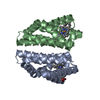 4zvaC 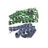 4zvbC 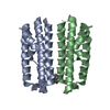 4zvcSC 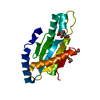 4zveC 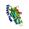 4zvfC 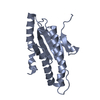 4zvgC 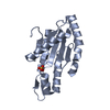 4zvhC S: Starting model for refinement C: citing same article ( |
|---|---|
| Similar structure data |

-Links
 PDBj
PDBj


- Assembly
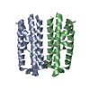
| Deposited unit | 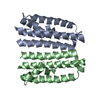
| ||||||||
|---|---|---|---|---|---|---|---|---|---|
| 1 |
| ||||||||
| Unit cell |
|
-Components
| #1: Protein | Mass: 14678.621 Da / Num. of mol.: 2 / Fragment: UNP residues 173-298 Source method: isolated from a genetically manipulated source Source: (gene. exp.) #2: Water | ChemComp-HOH / |  Mass: 18.015 Da / Num. of mol.: 62 / Source method: isolated from a natural source / Formula: H2O Mass: 18.015 Da / Num. of mol.: 62 / Source method: isolated from a natural source / Formula: H2O |
|---|
-Experimental details
-Experiment
| Experiment | Method: X-RAY DIFFRACTION |
|---|
- Sample preparation
| Crystal grow | Temperature: 293 K / Method: vapor diffusion Details: 0.2M ammonium dihydrogen phosphate, 20% (w/v) PEG 3350 |
|---|
-Data collection
| Diffraction | Mean temperature: 100 K |
|---|---|
| Diffraction source | Source: SYNCHROTRON / Site: SLS  / Beamline: X10SA / Wavelength: 0.99999 Å / Beamline: X10SA / Wavelength: 0.99999 Å |
| Detector | Type: DECTRIS PILATUS 6M / Detector: PIXEL / Date: Feb 25, 2014 |
| Radiation | Monochromator: Si(111) / Protocol: SINGLE WAVELENGTH / Monochromatic (M) / Laue (L): M / Scattering type: x-ray |
| Radiation wavelength | Wavelength: 0.99999 Å / Relative weight: 1 |
| Reflection | Resolution: 1.9→50 Å / Num. obs: 16326 / % possible obs: 100 % / Redundancy: 12.5 % / Rmerge(I) obs: 0.062 / Net I/σ(I): 25.22 |
| Reflection shell | Resolution: 1.9→2 Å / Redundancy: 13.2 % / Rmerge(I) obs: 0.624 / Mean I/σ(I) obs: 4.48 / % possible all: 100 |
- Processing
| Software |
| |||||||||||||||||||||||||||||||||||||||||||||||||
|---|---|---|---|---|---|---|---|---|---|---|---|---|---|---|---|---|---|---|---|---|---|---|---|---|---|---|---|---|---|---|---|---|---|---|---|---|---|---|---|---|---|---|---|---|---|---|---|---|---|---|
| Refinement | Method to determine structure: MOLECULAR REPLACEMENT Starting model: 4ZVC Resolution: 1.9→46.325 Å / SU ML: 0.27 / Cross valid method: FREE R-VALUE / σ(F): 1.37 / Phase error: 29.16 / Stereochemistry target values: ML
| |||||||||||||||||||||||||||||||||||||||||||||||||
| Solvent computation | Shrinkage radii: 0.9 Å / VDW probe radii: 1.11 Å / Solvent model: FLAT BULK SOLVENT MODEL | |||||||||||||||||||||||||||||||||||||||||||||||||
| Refinement step | Cycle: LAST / Resolution: 1.9→46.325 Å
| |||||||||||||||||||||||||||||||||||||||||||||||||
| Refine LS restraints |
| |||||||||||||||||||||||||||||||||||||||||||||||||
| LS refinement shell |
|
