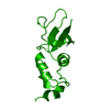
Entry Database : PDB / ID : 1s1gTitle Crystal Structure of Kv4.3 T1 Domain Potassium voltage-gated channel subfamily D member 3 Keywords / / / Function / homology Function Domain/homology Component
/ / / / / / / / / / / / / / / / / / / / / / / / / / / / / / / / / / / / / / / / / / / / / / / / / / / / / / / / / / / / / / / / Biological species Homo sapiens (human)Method / / / Resolution : 2.6 Å Authors Scannevin, R.H. / Wang, K.W. / Jow, F. / Megules, J. / Kopsco, D.C. / Edris, W. / Carroll, K.C. / Lu, Q. / Xu, W.X. / Xu, Z.B. ...Scannevin, R.H. / Wang, K.W. / Jow, F. / Megules, J. / Kopsco, D.C. / Edris, W. / Carroll, K.C. / Lu, Q. / Xu, W.X. / Xu, Z.B. / Katz, A.H. / Olland, S. / Lin, L. / Taylor, M. / Stahl, M. / Malakian, K. / Somers, W. / Mosyak, L. / Bowlby, M.R. / Chanda, P. / Rhodes, K.J. Journal : Neuron / Year : 2004Title : Two N-terminal domains of Kv4 K(+) channels regulate binding to and modulation by KChIP1.Authors: Scannevin, R.H. / Wang, K.W. / Jow, F. / Megules, J. / Kopsco, D.C. / Edris, W. / Carroll, K.C. / Xu, W.X. / Xu, Z.B. / Katz, A.H. / Olland, S. / Lin, L. / Taylor, M. / Stahl, M. / Malakian, ... Authors : Scannevin, R.H. / Wang, K.W. / Jow, F. / Megules, J. / Kopsco, D.C. / Edris, W. / Carroll, K.C. / Xu, W.X. / Xu, Z.B. / Katz, A.H. / Olland, S. / Lin, L. / Taylor, M. / Stahl, M. / Malakian, K. / Somers, W. / Mosyak, L. / Bowlby, M.R. / Chanda, P. / Rhodes, K.J. History Deposition Jan 6, 2004 Deposition site / Processing site Revision 1.0 Mar 23, 2004 Provider / Type Revision 1.1 Apr 29, 2008 Group Revision 1.2 Jul 13, 2011 Group / Version format complianceRevision 1.3 Jun 27, 2018 Group / Database references / Category / Item Revision 1.4 Aug 23, 2023 Group Data collection / Database references ... Data collection / Database references / Derived calculations / Refinement description Category chem_comp_atom / chem_comp_bond ... chem_comp_atom / chem_comp_bond / database_2 / pdbx_initial_refinement_model / pdbx_struct_conn_angle / struct_conn / struct_ref_seq_dif / struct_site Item _database_2.pdbx_DOI / _database_2.pdbx_database_accession ... _database_2.pdbx_DOI / _database_2.pdbx_database_accession / _pdbx_struct_conn_angle.ptnr1_auth_asym_id / _pdbx_struct_conn_angle.ptnr1_auth_comp_id / _pdbx_struct_conn_angle.ptnr1_auth_seq_id / _pdbx_struct_conn_angle.ptnr1_label_asym_id / _pdbx_struct_conn_angle.ptnr1_label_atom_id / _pdbx_struct_conn_angle.ptnr1_label_comp_id / _pdbx_struct_conn_angle.ptnr1_label_seq_id / _pdbx_struct_conn_angle.ptnr1_symmetry / _pdbx_struct_conn_angle.ptnr3_auth_asym_id / _pdbx_struct_conn_angle.ptnr3_auth_comp_id / _pdbx_struct_conn_angle.ptnr3_auth_seq_id / _pdbx_struct_conn_angle.ptnr3_label_asym_id / _pdbx_struct_conn_angle.ptnr3_label_atom_id / _pdbx_struct_conn_angle.ptnr3_label_comp_id / _pdbx_struct_conn_angle.ptnr3_label_seq_id / _pdbx_struct_conn_angle.ptnr3_symmetry / _pdbx_struct_conn_angle.value / _struct_conn.pdbx_dist_value / _struct_conn.ptnr1_auth_asym_id / _struct_conn.ptnr1_auth_comp_id / _struct_conn.ptnr1_auth_seq_id / _struct_conn.ptnr1_label_asym_id / _struct_conn.ptnr1_label_atom_id / _struct_conn.ptnr1_label_comp_id / _struct_conn.ptnr1_label_seq_id / _struct_conn.ptnr1_symmetry / _struct_conn.ptnr2_auth_asym_id / _struct_conn.ptnr2_auth_comp_id / _struct_conn.ptnr2_auth_seq_id / _struct_conn.ptnr2_label_asym_id / _struct_conn.ptnr2_label_atom_id / _struct_conn.ptnr2_label_comp_id / _struct_conn.ptnr2_label_seq_id / _struct_conn.ptnr2_symmetry / _struct_ref_seq_dif.details / _struct_site.pdbx_auth_asym_id / _struct_site.pdbx_auth_comp_id / _struct_site.pdbx_auth_seq_id
Show all Show less
 Movie
Movie Controller


 Open data
 Basic information
 Components
 Keywords
 Function and homology information
 Homo sapiens (human)
 X-RAY DIFFRACTION /
  Authors
 Citation
 Structure visualization
 Downloads & links
 Other downloads


 PDBj
PDBj


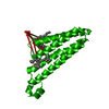
 Assembly
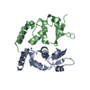


 Mass: 65.409 Da / Num. of mol.: 2 / Source method: obtained synthetically / Formula: Zn
 Mass: 18.015 Da / Num. of mol.: 28 / Source method: isolated from a natural source / Formula: H2O
 Sample preparation
 / Beamline: 5.0.2 / Wavelength: 1.1 Å
 Processing