 Movie
Movie Controller

[English] 日本語
 Yorodumi
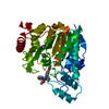
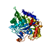
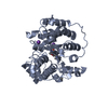
- PDB-6uo3: Crystal structure of Danio rerio histone deacetylase 6 catalytic ... -
+ Open data

- Basic information
| Entry | Database: PDB / ID: 6uo3 | ||||||
|---|---|---|---|---|---|---|---|
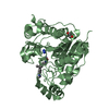
| Title | Crystal structure of Danio rerio histone deacetylase 6 catalytic domain 1 (CD1) complexed with AR-42 | ||||||
 Components Components | HDAC6 | ||||||
 Keywords Keywords | Hydrolase/Hydrolase Inhibitor / Histone Deacetylase / Hydrolase-Hydrolase Inhibitor complex | ||||||
| Function / homology |  Function and homology information Function and homology informationAggrephagy / negative regulation of cellular component organization / positive regulation of cellular component organization / deacetylase activity / tubulin deacetylase activity / mitochondrion localization / definitive hemopoiesis / regulation of microtubule-based process / protein lysine deacetylase activity / potassium ion binding ...Aggrephagy / negative regulation of cellular component organization / positive regulation of cellular component organization / deacetylase activity / tubulin deacetylase activity / mitochondrion localization / definitive hemopoiesis / regulation of microtubule-based process / protein lysine deacetylase activity / potassium ion binding / response to stress / hematopoietic progenitor cell differentiation / swimming behavior / transferase activity / chromatin organization / actin binding / angiogenesis / perikaryon / axon / centrosome / dendrite / zinc ion binding / nucleus / cytosol / cytoplasm Similarity search - Function | ||||||
| Biological species |  | ||||||
| Method |  X-RAY DIFFRACTION / SYNCHROTRON / MOLECULAR REPLACEMENT / Resolution: 1.09000122285 Å X-RAY DIFFRACTION / SYNCHROTRON / MOLECULAR REPLACEMENT / Resolution: 1.09000122285 Å | ||||||
 Authors Authors | Osko, J.D. / Christianson, D.W. | ||||||
| Funding support |  United States, 1items United States, 1items
| ||||||
 Citation Citation | Journal: Biochemistry / Year: 2019 Title: Structural Basis of Catalysis and Inhibition of HDAC6 CD1, the Enigmatic Catalytic Domain of Histone Deacetylase 6. Authors: Osko, J.D. / Christianson, D.W. | ||||||
| History |
|
- Structure visualization
| Structure viewer | Molecule: MolmilJmol/JSmol |
|---|
- Downloads & links
-Download
| PDBx/mmCIF format | 6uo3.cif.gz | 122.2 KB | Display | PDBx/mmCIF format |
|---|---|---|---|---|
| PDB format | pdb6uo3.ent.gz | 72.8 KB | Display | PDB format |
| PDBx/mmJSON format | 6uo3.json.gz | Tree view | PDBx/mmJSON format | |
| Others |  Other downloads Other downloads |
-Validation report
| Arichive directory | https://data.pdbj.org/pub/pdb/validation_reports/uo/6uo3ftp://data.pdbj.org/pub/pdb/validation_reports/uo/6uo3 | HTTPS FTP |
|---|
-Related structure data
| Related structure data | 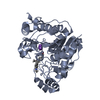 6uo2C  6uo4C  6uo5C  6uo7C  6uobC  6uocC 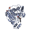 5eefS S: Starting model for refinement C: citing same article ( |
|---|---|
| Similar structure data |


-Links
 PDBj
PDBj- Assembly
| Deposited unit | 
| ||||||||||||
|---|---|---|---|---|---|---|---|---|---|---|---|---|---|
| 1 |
| ||||||||||||
| Unit cell |
|
-Components
-Protein , 1 types, 1 molecules A
| #1: Protein | Mass: 40294.594 Da / Num. of mol.: 1 Source method: isolated from a genetically manipulated source Source: (gene. exp.)  |
|---|
-Non-polymers , 5 types, 420 molecules 


| #2: Chemical | ChemComp-ZN /  Mass: 65.409 Da / Num. of mol.: 1 / Source method: obtained synthetically / Formula: Zn Mass: 65.409 Da / Num. of mol.: 1 / Source method: obtained synthetically / Formula: Zn | ||||||
|---|---|---|---|---|---|---|---|
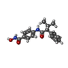
| #3: Chemical |  Mass: 39.098 Da / Num. of mol.: 2 / Source method: obtained synthetically / Formula: K Mass: 39.098 Da / Num. of mol.: 2 / Source method: obtained synthetically / Formula: K#4: Chemical | ChemComp-QCP / |  Mass: 312.363 Da / Num. of mol.: 1 / Source method: obtained synthetically / Formula: C18H20N2O3 / Feature type: SUBJECT OF INVESTIGATION / Comment: inhibitor*YM Mass: 312.363 Da / Num. of mol.: 1 / Source method: obtained synthetically / Formula: C18H20N2O3 / Feature type: SUBJECT OF INVESTIGATION / Comment: inhibitor*YM#5: Chemical |  Mass: 62.068 Da / Num. of mol.: 3 / Source method: obtained synthetically / Formula: C2H6O2 Mass: 62.068 Da / Num. of mol.: 3 / Source method: obtained synthetically / Formula: C2H6O2#6: Water | ChemComp-HOH / | Mass: 18.015 Da / Num. of mol.: 413 / Source method: isolated from a natural source / Formula: H2O |
-Details
| Has ligand of interest | Y |
|---|
-Experimental details
-Experiment
| Experiment | Method: X-RAY DIFFRACTION / Number of used crystals: 1 |
|---|
- Sample preparation
| Crystal | Density Matthews: 2.52 Å3/Da / Density % sol: 51.25 % |
|---|---|
| Crystal grow | Temperature: 277 K / Method: vapor diffusion, sitting drop Details: 10 mg/ml HDAC6 CD1 2 mM Inhibitor 0.2 M ammonium tartrate dibasic (pH 7.0) 20% PEG 3350 1:1 ratio protein to precipitant |
-Data collection
| Diffraction | Mean temperature: 100 K / Serial crystal experiment: N |
|---|---|
| Diffraction source | Source: SYNCHROTRON / Site: APS / Beamline: 24-ID-C / Wavelength: 0.98 Å |
| Detector | Type: DECTRIS PILATUS 6M-F / Detector: PIXEL / Date: Aug 2, 2019 |
| Radiation | Protocol: SINGLE WAVELENGTH / Monochromatic (M) / Laue (L): M / Scattering type: x-ray |
| Radiation wavelength | Wavelength: 0.98 Å / Relative weight: 1 |
| Reflection | Resolution: 1.09→43.0926147252 Å / Num. obs: 169488 / % possible obs: 99.7 % / Redundancy: 6 % / Biso Wilson estimate: 9.50667468151 Å2 / CC1/2: 0.986 / Rmerge(I) obs: 0.14 / Rpim(I) all: 0.062 / Net I/σ(I): 7.2 |
| Reflection shell | Resolution: 1.09→1.12 Å / Rmerge(I) obs: 0.995 / Mean I/σ(I) obs: 2 / Num. unique obs: 16796 / CC1/2: 0.776 / Rpim(I) all: 0.445 |
- Processing
| Software |
| |||||||||||||||||||||||||||||||||||||||||||||||||||||||||||||||||||||||||||||||||||||||||||||||||||||||||||||||||||||||||||||||||||||||||||||||||||||||||||||||||||||||||||||||||||||||||||||||||||||||||||||||||||||||||
|---|---|---|---|---|---|---|---|---|---|---|---|---|---|---|---|---|---|---|---|---|---|---|---|---|---|---|---|---|---|---|---|---|---|---|---|---|---|---|---|---|---|---|---|---|---|---|---|---|---|---|---|---|---|---|---|---|---|---|---|---|---|---|---|---|---|---|---|---|---|---|---|---|---|---|---|---|---|---|---|---|---|---|---|---|---|---|---|---|---|---|---|---|---|---|---|---|---|---|---|---|---|---|---|---|---|---|---|---|---|---|---|---|---|---|---|---|---|---|---|---|---|---|---|---|---|---|---|---|---|---|---|---|---|---|---|---|---|---|---|---|---|---|---|---|---|---|---|---|---|---|---|---|---|---|---|---|---|---|---|---|---|---|---|---|---|---|---|---|---|---|---|---|---|---|---|---|---|---|---|---|---|---|---|---|---|---|---|---|---|---|---|---|---|---|---|---|---|---|---|---|---|---|---|---|---|---|---|---|---|---|---|---|---|---|---|---|---|---|
| Refinement | Method to determine structure: MOLECULAR REPLACEMENT Starting model: 5EEF Resolution: 1.09000122285→43.0926147252 Å / SU ML: 0.100731551156 / Cross valid method: FREE R-VALUE / σ(F): 1.33645998405 / Phase error: 19.8872764084
| |||||||||||||||||||||||||||||||||||||||||||||||||||||||||||||||||||||||||||||||||||||||||||||||||||||||||||||||||||||||||||||||||||||||||||||||||||||||||||||||||||||||||||||||||||||||||||||||||||||||||||||||||||||||||
| Solvent computation | Shrinkage radii: 0.9 Å / VDW probe radii: 1.11 Å | |||||||||||||||||||||||||||||||||||||||||||||||||||||||||||||||||||||||||||||||||||||||||||||||||||||||||||||||||||||||||||||||||||||||||||||||||||||||||||||||||||||||||||||||||||||||||||||||||||||||||||||||||||||||||
| Displacement parameters | Biso mean: 14.2140592905 Å2 | |||||||||||||||||||||||||||||||||||||||||||||||||||||||||||||||||||||||||||||||||||||||||||||||||||||||||||||||||||||||||||||||||||||||||||||||||||||||||||||||||||||||||||||||||||||||||||||||||||||||||||||||||||||||||
| Refinement step | Cycle: LAST / Resolution: 1.09000122285→43.0926147252 Å
| |||||||||||||||||||||||||||||||||||||||||||||||||||||||||||||||||||||||||||||||||||||||||||||||||||||||||||||||||||||||||||||||||||||||||||||||||||||||||||||||||||||||||||||||||||||||||||||||||||||||||||||||||||||||||
| Refine LS restraints |
| |||||||||||||||||||||||||||||||||||||||||||||||||||||||||||||||||||||||||||||||||||||||||||||||||||||||||||||||||||||||||||||||||||||||||||||||||||||||||||||||||||||||||||||||||||||||||||||||||||||||||||||||||||||||||
| LS refinement shell |
|
