 Movie
Movie Controller

[English] 日本語
 Yorodumi
- PDB-4zq0: crystal structure of Giardia 14-3-3 in complex with the phosphope... -
+ Open data

- Basic information
| Entry | Database: PDB / ID: 4zq0 | ||||||
|---|---|---|---|---|---|---|---|
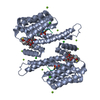
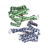
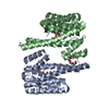
| Title | crystal structure of Giardia 14-3-3 in complex with the phosphopeptide A8Ap | ||||||
 Components Components |
| ||||||
 Keywords Keywords | SIGNALING PROTEIN / phosphopeptide binding protein-protein interaction | ||||||
| Function / homology |  Function and homology information Function and homology information | ||||||
| Biological species |  Giardia lamblia P15 (eukaryote) Giardia lamblia P15 (eukaryote)synthetic construct (others) | ||||||
| Method |  X-RAY DIFFRACTION / SYNCHROTRON / MOLECULAR REPLACEMENT / molecular replacement / Resolution: 3.1 Å X-RAY DIFFRACTION / SYNCHROTRON / MOLECULAR REPLACEMENT / molecular replacement / Resolution: 3.1 Å | ||||||
 Authors Authors | Fiorillo, A. / Ilari, A. | ||||||
 Citation Citation | Journal: J.Chem.Inf.Model. / Year: 2015 Title: Molecular Dynamics Simulations and Structural Analysis of Giardia duodenalis 14-3-3 Protein-Protein Interactions. Authors: Cau, Y. / Fiorillo, A. / Mori, M. / Ilari, A. / Botta, M. / Lalle, M. #1: Journal: Plos One / Year: 2014Title: The crystal structure of Giardia duodenalis 14-3-3 in the apo form: when protein post-translational modifications make the difference. Authors: Fiorillo, A. / di Marino, D. / Bertuccini, L. / Via, A. / Pozio, E. / Camerini, S. / Ilari, A. / Lalle, M. | ||||||
| History |
|
- Structure visualization
| Structure viewer | Molecule: MolmilJmol/JSmol |
|---|
- Downloads & links
-Download
| PDBx/mmCIF format | 4zq0.cif.gz | 195.7 KB | Display | PDBx/mmCIF format |
|---|---|---|---|---|
| PDB format | pdb4zq0.ent.gz | 157.9 KB | Display | PDB format |
| PDBx/mmJSON format | 4zq0.json.gz | Tree view | PDBx/mmJSON format | |
| Others |  Other downloads Other downloads |
-Validation report
| Arichive directory | https://data.pdbj.org/pub/pdb/validation_reports/zq/4zq0ftp://data.pdbj.org/pub/pdb/validation_reports/zq/4zq0 | HTTPS FTP |
|---|
-Related structure data
| Related structure data |  5by9C  4f7rS S: Starting model for refinement C: citing same article ( |
|---|---|
| Similar structure data |


-Links
 PDBj
PDBj
- Assembly
| Deposited unit | 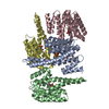
| |||||||||||||||||||||||||||||||||||||||||||||||||||||||||||||||||||||||||||||||||||||||||||||||||||||||||||||||||||||||||||||||||||||||||||||||||||||||||
|---|---|---|---|---|---|---|---|---|---|---|---|---|---|---|---|---|---|---|---|---|---|---|---|---|---|---|---|---|---|---|---|---|---|---|---|---|---|---|---|---|---|---|---|---|---|---|---|---|---|---|---|---|---|---|---|---|---|---|---|---|---|---|---|---|---|---|---|---|---|---|---|---|---|---|---|---|---|---|---|---|---|---|---|---|---|---|---|---|---|---|---|---|---|---|---|---|---|---|---|---|---|---|---|---|---|---|---|---|---|---|---|---|---|---|---|---|---|---|---|---|---|---|---|---|---|---|---|---|---|---|---|---|---|---|---|---|---|---|---|---|---|---|---|---|---|---|---|---|---|---|---|---|---|---|
| 1 | 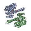
| |||||||||||||||||||||||||||||||||||||||||||||||||||||||||||||||||||||||||||||||||||||||||||||||||||||||||||||||||||||||||||||||||||||||||||||||||||||||||
| 2 | 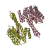
| |||||||||||||||||||||||||||||||||||||||||||||||||||||||||||||||||||||||||||||||||||||||||||||||||||||||||||||||||||||||||||||||||||||||||||||||||||||||||
| Unit cell |
| |||||||||||||||||||||||||||||||||||||||||||||||||||||||||||||||||||||||||||||||||||||||||||||||||||||||||||||||||||||||||||||||||||||||||||||||||||||||||
| Noncrystallographic symmetry (NCS) | NCS domain:
NCS domain segments: Ens-ID: 1 / Refine code: 4
|