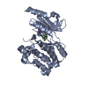
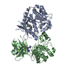
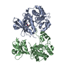
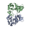
[English] 日本語
Yorodumi
- PDB-4zlo: Serine/threonine-protein kinase PAK1 complexed with a dibenzodiaz... -

+
Open data


ID or keywords:

Loading...

-
Basic information

Entry
Database: PDB / ID: 4zlo
TitleSerine/threonine-protein kinase PAK1 complexed with a dibenzodiazepine: identification of an allosteric site on PAK1
ComponentsSerine/threonine-protein kinase PAK 1
KeywordsTransferase/transferase inhibitor / dibenzodazepine / PAK1 / kinase / allosteric inhibitor / Transferase-transferase inhibitor complex
Function / homology
Function and homology information


negative regulation of cell proliferation involved in contact inhibition / protein localization to cytoplasmic stress granule / positive regulation of microtubule nucleation / hepatocyte growth factor receptor signaling pathway / RHO GTPases Activate ROCKs / gamma-tubulin binding / Activation of RAC1 / CD28 dependent Vav1 pathway / positive regulation of intracellular estrogen receptor signaling pathway / Ephrin signaling ...negative regulation of cell proliferation involved in contact inhibition / protein localization to cytoplasmic stress granule / positive regulation of microtubule nucleation / hepatocyte growth factor receptor signaling pathway / RHO GTPases Activate ROCKs / gamma-tubulin binding / Activation of RAC1 / CD28 dependent Vav1 pathway / positive regulation of intracellular estrogen receptor signaling pathway / Ephrin signaling / RHOV GTPase cycle / DSCAM interactions / regulation of axonogenesis / branching morphogenesis of an epithelial tube / Fc-gamma receptor signaling pathway involved in phagocytosis / RHOJ GTPase cycle / exocytosis / RHOQ GTPase cycle / RHO GTPases activate PAKs / stimulatory C-type lectin receptor signaling pathway / regulation of MAPK cascade / CDC42 GTPase cycle / RHOH GTPase cycle / RHOU GTPase cycle / Sema3A PAK dependent Axon repulsion / Generation of second messenger molecules / intercalated disc / Smooth Muscle Contraction / localization / ephrin receptor signaling pathway / RAC2 GTPase cycle / RAC3 GTPase cycle / positive regulation of JUN kinase activity / RHO GTPases activate PKNs / positive regulation of microtubule polymerization / positive regulation of stress fiber assembly / ruffle / collagen binding / RAC1 GTPase cycle / EPHB-mediated forward signaling / CD209 (DC-SIGN) signaling / neuron projection morphogenesis / VEGFR2 mediated vascular permeability / Signal transduction by L1 / actin filament / regulation of actin cytoskeleton organization / FCERI mediated MAPK activation / MAPK6/MAPK4 signaling / wound healing / G beta:gamma signalling through CDC42 / Regulation of actin dynamics for phagocytic cup formation / ruffle membrane / Z disc / cell-cell junction / cell migration / lamellipodium / positive regulation of peptidyl-serine phosphorylation / chromosome / actin cytoskeleton organization / nuclear membrane / protein autophosphorylation / non-specific serine/threonine protein kinase / protein kinase activity / intracellular signal transduction / positive regulation of cell migration / chromatin remodeling / positive regulation of protein phosphorylation / phosphorylation / axon / protein phosphorylation / focal adhesion / protein serine kinase activity / protein serine/threonine kinase activity / centrosome / apoptotic process / DNA damage response / dendrite / positive regulation of cell population proliferation / protein-containing complex / nucleoplasm / ATP binding / identical protein binding / plasma membrane / cytosol / cytoplasm
Similarity search - Function
p21 activated kinase binding domain / CRIB domain superfamily / P21-Rho-binding domain / CRIB domain profile. / P21-Rho-binding domain / CRIB domain / Transferase(Phosphotransferase) domain 1 / Transferase(Phosphotransferase); domain 1 / Phosphorylase Kinase; domain 1 / Phosphorylase Kinase; domain 1 ...p21 activated kinase binding domain / CRIB domain superfamily / P21-Rho-binding domain / CRIB domain profile. / P21-Rho-binding domain / CRIB domain / Transferase(Phosphotransferase) domain 1 / Transferase(Phosphotransferase); domain 1 / Phosphorylase Kinase; domain 1 / Phosphorylase Kinase; domain 1 / Serine/threonine-protein kinase, active site / Serine/Threonine protein kinases active-site signature. / Protein kinase domain / Serine/Threonine protein kinases, catalytic domain / Protein kinase, ATP binding site / Protein kinases ATP-binding region signature. / Protein kinase domain profile. / Protein kinase domain / Protein kinase-like domain superfamily / 2-Layer Sandwich / Orthogonal Bundle / Mainly Alpha / Alpha Beta
Similarity search - Domain/homology
Chem-4PV / Serine/threonine-protein kinase PAK 1
Similarity search - Component
Biological speciesHomo sapiens (human)
MethodX-RAY DIFFRACTION / Resolution: 2.5 Å
AuthorsBellamacina, C.R. / Bussiere, D.E.
CitationJournal: Acs Med.Chem.Lett. / Year: 2015
Title: Optimization of a Dibenzodiazepine Hit to a Potent and Selective Allosteric PAK1 Inhibitor.
Authors: Karpov, A.S. / Amiri, P. / Bellamacina, C. / Bellance, M.H. / Breitenstein, W. / Daniel, D. / Denay, R. / Fabbro, D. / Fernandez, C. / Galuba, I. / Guerro-Lagasse, S. / Gutmann, S. / Hinh, L. ...Authors: Karpov, A.S. / Amiri, P. / Bellamacina, C. / Bellance, M.H. / Breitenstein, W. / Daniel, D. / Denay, R. / Fabbro, D. / Fernandez, C. / Galuba, I. / Guerro-Lagasse, S. / Gutmann, S. / Hinh, L. / Jahnke, W. / Klopp, J. / Lai, A. / Lindvall, M.K. / Ma, S. / Mobitz, H. / Pecchi, S. / Rummel, G. / Shoemaker, K. / Trappe, J. / Voliva, C. / Cowan-Jacob, S.W. / Marzinzik, A.L.
History
DepositionMay 1, 2015Deposition site: RCSB / Processing site: RCSB
Revision 1.0Aug 5, 2015Provider: repository / Type: Initial release

-
Structure visualization

Structure viewerMolecule:
MolmilJmol/JSmol

Downloads & links

-
Assembly

Deposited unit
A: Serine/threonine-protein kinase PAK 1
B: Serine/threonine-protein kinase PAK 1
hetero molecules


Theoretical massNumber of molelcules
Total (without water)67,0994
Polymers66,6782
Non-polymers4202
Water2,180121
1
A: Serine/threonine-protein kinase PAK 1
hetero molecules

B: Serine/threonine-protein kinase PAK 1


Theoretical massNumber of molelcules
Total (without water)67,0994
Polymers66,6782
Non-polymers4202
Water362
TypeNameSymmetry operationNumber
identity operation1_555x,y,z1
crystal symmetry operation1_554x,y,z-11
Buried area1520 Å2
ΔGint-9 kcal/mol
Surface area25640 Å2
MethodPISA
2


  • Idetical with deposited unit
  • defined by author&software
TypeNameSymmetry operationNumber
identity operation1_555x,y,z1
Buried area950 Å2
ΔGint-11 kcal/mol
Surface area26210 Å2
MethodPISA
Unit cell
Length a, b, c (Å)63.514, 79.340, 65.789
Angle α, β, γ (deg.)90.00, 108.11, 90.00
Int Tables number4
Space group name H-MP1211

-
Components

#1: Protein Serine/threonine-protein kinase PAK 1 / Alpha-PAK / p21-activated kinase 1 / PAK-1 / p65-PAK


Mass: 33339.227 Da / Num. of mol.: 2
Source method: isolated from a genetically manipulated source
Details: Residue 177 is phosphorylated Threonine / Source: (gene. exp.) Homo sapiens (human) / Gene: PAK1 / Production host: Escherichia coli (E. coli)
References: UniProt: Q13153, non-specific serine/threonine protein kinase
#2: Chemical ChemComp-4PV / 2,8-difluoro-11-(4-methylpiperazin-1-yl)-5H-dibenzo[b,e][1,4]diazepine


Mass: 328.359 Da / Num. of mol.: 1 / Source method: obtained synthetically / Formula: C18H18F2N4
#3: Chemical ChemComp-GOL / GLYCEROL / GLYCERIN / PROPANE-1,2,3-TRIOL


Mass: 92.094 Da / Num. of mol.: 1 / Source method: obtained synthetically / Formula: C3H8O3
#4: Water ChemComp-HOH / water


Mass: 18.015 Da / Num. of mol.: 121 / Source method: isolated from a natural source / Formula: H2O

-
Experimental details

-
Experiment

ExperimentMethod: X-RAY DIFFRACTION

-
Sample preparation

CrystalDensity Matthews: 2.36 Å3/Da / Density % sol: 47.94 %
Crystal growTemperature: 291 K / Method: vapor diffusion, hanging drop / pH: 6.5
Details: 22-27% PEG3350, 500-700MM NACL, 100MM BIS-TRIS, PH 6.5

-
Data collection

DiffractionMean temperature: 100 K / Ambient temp details: cryo conditions
Diffraction sourceSource: ROTATING ANODE / Type: RIGAKU FR-E SUPERBRIGHT / Wavelength: 1.54178 Å
DetectorType: RIGAKU RAXIS HTC / Detector: IMAGE PLATE / Date: Jan 7, 2009
RadiationProtocol: SINGLE WAVELENGTH / Monochromatic (M) / Laue (L): M / Scattering type: x-ray
Radiation wavelengthWavelength: 1.54178 Å / Relative weight: 1
ReflectionResolution: 2.5→62.5 Å / Num. all: 21368 / Num. obs: 21368 / % possible obs: 98.8 % / Redundancy: 3.2 % / Biso Wilson estimate: 64.1 Å2 / Net I/σ(I): 13.3
Reflection shellResolution: 2.5→2.77 Å / Redundancy: 2.9 % / Rmerge(I) obs: 0.415 / Mean I/σ(I) obs: 2.6 / % possible all: 96

-
Processing

Software
NameVersionClassification
BUSTER2.11.4refinement
MOSFLMdata reduction
SCALAdata scaling
REFMACphasing
RefinementResolution: 2.5→52.31 Å / Cor.coef. Fo:Fc: 0.935 / Cor.coef. Fo:Fc free: 0.9161 / SU R Cruickshank DPI: 0.477 / Cross valid method: THROUGHOUT / σ(F): 0 / SU R Blow DPI: 0.459 / SU Rfree Blow DPI: 0.243 / SU Rfree Cruickshank DPI: 0.248
RfactorNum. reflection% reflectionSelection details
Rfree0.2224 1081 5.06 %RANDOM
Rwork0.1946 ---
obs0.196 21368 98.8 %-
Displacement parametersBiso mean: 62.7 Å2
Baniso -1Baniso -2Baniso -3
1--2.5514 Å20 Å2-7.5359 Å2
2---1.9211 Å20 Å2
3---4.4725 Å2
Refine analyzeLuzzati coordinate error obs: 0.315 Å
Refinement stepCycle: 1 / Resolution: 2.5→52.31 Å
ProteinNucleic acidLigandSolventTotal
Num. atoms4066 0 30 121 4217
Refine LS restraints
Refine-IDTypeDev idealNumberRestraint functionWeight
X-RAY DIFFRACTIONt_bond_d0.014182HARMONIC2
X-RAY DIFFRACTIONt_angle_deg1.135674HARMONIC2
X-RAY DIFFRACTIONt_dihedral_angle_d1429SINUSOIDAL2
X-RAY DIFFRACTIONt_incorr_chiral_ct
X-RAY DIFFRACTIONt_pseud_angle
X-RAY DIFFRACTIONt_trig_c_planes94HARMONIC2
X-RAY DIFFRACTIONt_gen_planes616HARMONIC5
X-RAY DIFFRACTIONt_it4182HARMONIC20
X-RAY DIFFRACTIONt_nbd
X-RAY DIFFRACTIONt_omega_torsion2.82
X-RAY DIFFRACTIONt_other_torsion18.01
X-RAY DIFFRACTIONt_improper_torsion
X-RAY DIFFRACTIONt_chiral_improper_torsion581SEMIHARMONIC5
X-RAY DIFFRACTIONt_sum_occupancies1HARMONIC1
X-RAY DIFFRACTIONt_utility_distance
X-RAY DIFFRACTIONt_utility_angle
X-RAY DIFFRACTIONt_utility_torsion
X-RAY DIFFRACTIONt_ideal_dist_contact4808SEMIHARMONIC4
LS refinement shellResolution: 2.5→2.62 Å / Total num. of bins used: 11
RfactorNum. reflection% reflection
Rfree0.28 123 4.61 %
Rwork0.2094 2545 -
all0.2125 2668 -
obs--98.8 %
Refinement TLS params.

Method: refined / Refine-ID: X-RAY DIFFRACTION

IDL112)L122)L132)L222)L232)L332)S11 (Å °)S12 (Å °)S13 (Å °)S21 (Å °)S22 (Å °)S23 (Å °)S31 (Å °)S32 (Å °)S33 (Å °)T112)T122)T132)T222)T232)T332)Origin x (Å)Origin y (Å)Origin z (Å)
11.3221-0.6516-0.61352.0107-0.44072.08640.01820.22340.0221-0.1094-0.0894-0.15820.1047-0.07060.0712-0.1117-0.0430.011-0.11620.0036-0.113932.11595.1771-14.5141
23.3059-0.3145-1.2152.6534-0.52033.0827-0.17950.1178-0.32180.02550.00120.2681-0.0143-0.41360.1783-0.1796-0.06610.0196-0.1233-0.0205-0.2129-1.83210.976113.3558
Refinement TLS group
IDRefine-IDRefine TLS-IDSelection details
1X-RAY DIFFRACTION1{ A|* }
2X-RAY DIFFRACTION2{ B|* }

+
About Yorodumi

-
News

-
Feb 9, 2022. New format data for meta-information of EMDB entries

New format data for meta-information of EMDB entries

  • Version 3 of the EMDB header file is now the official format.
  • The previous official version 1.9 will be removed from the archive.

Related info.:EMDB header

External links:wwPDB to switch to version 3 of the EMDB data model

-
Aug 12, 2020. Covid-19 info

Covid-19 info

URL: https://pdbjlvh1.pdbj.org/emnavi/covid19.php

New page: Covid-19 featured information page in EM Navigator.

Related info.:Covid-19 info / Mar 5, 2020. Novel coronavirus structure data

+
Mar 5, 2020. Novel coronavirus structure data

Novel coronavirus structure data

Related info.:Yorodumi Speices / Aug 12, 2020. Covid-19 info

External links:COVID-19 featured content - PDBj / Molecule of the Month (242):Coronavirus Proteases

+
Jan 31, 2019. EMDB accession codes are about to change! (news from PDBe EMDB page)

EMDB accession codes are about to change! (news from PDBe EMDB page)

  • The allocation of 4 digits for EMDB accession codes will soon come to an end. Whilst these codes will remain in use, new EMDB accession codes will include an additional digit and will expand incrementally as the available range of codes is exhausted. The current 4-digit format prefixed with “EMD-” (i.e. EMD-XXXX) will advance to a 5-digit format (i.e. EMD-XXXXX), and so on. It is currently estimated that the 4-digit codes will be depleted around Spring 2019, at which point the 5-digit format will come into force.
  • The EM Navigator/Yorodumi systems omit the EMD- prefix.

Related info.:Q: What is EMD? / ID/Accession-code notation in Yorodumi/EM Navigator

External links:EMDB Accession Codes are Changing Soon! / Contact to PDBj

+
Jul 12, 2017. Major update of PDB

Major update of PDB

  • wwPDB released updated PDB data conforming to the new PDBx/mmCIF dictionary.
  • This is a major update changing the version number from 4 to 5, and with Remediation, in which all the entries are updated.
  • In this update, many items about electron microscopy experimental information are reorganized (e.g. em_software).
  • Now, EM Navigator and Yorodumi are based on the updated data.

External links:wwPDB Remediation / Enriched Model Files Conforming to OneDep Data Standards Now Available in the PDB FTP Archive

-
Yorodumi

Thousand views of thousand structures

  • Yorodumi is a browser for structure data from EMDB, PDB, SASBDB, etc.
  • This page is also the successor to EM Navigator detail page, and also detail information page/front-end page for Omokage search.
  • The word "yorodu" (or yorozu) is an old Japanese word meaning "ten thousand". "mi" (miru) is to see.

Related info.:EMDB / PDB / SASBDB / Comparison of 3 databanks / Yorodumi Search / Aug 31, 2016. New EM Navigator & Yorodumi / Yorodumi Papers / Jmol/JSmol / Function and homology information / Changes in new EM Navigator and Yorodumi

Read more