 Movie
Movie Controller

[English] 日本語
 Yorodumi
- PDB-4zgo: Structure of C-terminally truncated Cdc123 from Schizosaccharomyc... -
+ Open data

- Basic information
| Entry | Database: PDB / ID: 4zgo | |||||||||
|---|---|---|---|---|---|---|---|---|---|---|
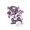
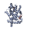
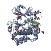
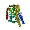
| Title | Structure of C-terminally truncated Cdc123 from Schizosaccharomyces pombe | |||||||||
 Components Components | Cell division cycle protein 123 | |||||||||
 Keywords Keywords | CELL CYCLE / ATP-grap fold / eIF2 assembly | |||||||||
| Function / homology | eukaryotic translation initiation factor 2 complex assembly / Cell division cycle protein 123 / D123 / protein folding chaperone / magnesium ion binding / ATP binding / cytoplasm / cytosol / Translation initiation factor eIF2 assembly protein Function and homology information Function and homology information | |||||||||
| Biological species |  | |||||||||
| Method |  X-RAY DIFFRACTION / SYNCHROTRON / MOLECULAR REPLACEMENT / Resolution: 2.063 Å X-RAY DIFFRACTION / SYNCHROTRON / MOLECULAR REPLACEMENT / Resolution: 2.063 Å | |||||||||
 Authors Authors | Panvert, M. / Dubiez, E. / Arnold, L. / Perez, J. / Seufert, W. / Mechulam, Y. / Schmitt, E. | |||||||||
| Funding support |  France, 2items France, 2items
| |||||||||
 Citation Citation | Journal: Structure / Year: 2015 Title: Cdc123, a Cell Cycle Regulator Needed for eIF2 Assembly, Is an ATP-Grasp Protein with Unique Features. Authors: Panvert, M. / Dubiez, E. / Arnold, L. / Perez, J. / Mechulam, Y. / Seufert, W. / Schmitt, E. | |||||||||
| History |
|
- Structure visualization
| Structure viewer | Molecule: MolmilJmol/JSmol |
|---|
- Downloads & links
-Download
| PDBx/mmCIF format | 4zgo.cif.gz | 121.4 KB | Display | PDBx/mmCIF format |
|---|---|---|---|---|
| PDB format | pdb4zgo.ent.gz | 92.9 KB | Display | PDB format |
| PDBx/mmJSON format | 4zgo.json.gz | Tree view | PDBx/mmJSON format | |
| Others |  Other downloads Other downloads |
-Validation report
| Arichive directory | https://data.pdbj.org/pub/pdb/validation_reports/zg/4zgoftp://data.pdbj.org/pub/pdb/validation_reports/zg/4zgo | HTTPS FTP |
|---|
-Related structure data


-Links
 PDBj
PDBj- Assembly
| Deposited unit | 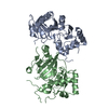
| ||||||||
|---|---|---|---|---|---|---|---|---|---|
| 1 | 
| ||||||||
| 2 | 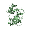
| ||||||||
| Unit cell |
|
-Components
| #1: Protein | Mass: 39110.246 Da / Num. of mol.: 2 / Fragment: unp residues 21-389 Source method: isolated from a genetically manipulated source Source: (gene. exp.) Gene: cdc123, SPAP27G11.03 / Plasmid: pET15b / Production host:  #2: Water | ChemComp-HOH / |  Mass: 18.015 Da / Num. of mol.: 198 / Source method: isolated from a natural source / Formula: H2O Mass: 18.015 Da / Num. of mol.: 198 / Source method: isolated from a natural source / Formula: H2O |
|---|
-Experimental details
-Experiment
| Experiment | Method: X-RAY DIFFRACTION |
|---|
- Sample preparation
| Crystal | Density Matthews: 2.16 Å3/Da / Density % sol: 43.05 % |
|---|---|
| Crystal grow | Temperature: 298 K / Method: vapor diffusion, sitting drop / pH: 5 / Details: 12% PEG3350, 4% tacsimate |
-Data collection
| Diffraction | Mean temperature: 100 K |
|---|---|
| Diffraction source | Source: SYNCHROTRON / Site: SOLEIL / Beamline: PROXIMA 1 / Wavelength: 0.979 Å |
| Detector | Type: PSI PILATUS 6M / Detector: PIXEL / Date: Jul 3, 2014 |
| Radiation | Protocol: SINGLE WAVELENGTH / Monochromatic (M) / Laue (L): M / Scattering type: x-ray |
| Radiation wavelength | Wavelength: 0.979 Å / Relative weight: 1 |
| Reflection | Resolution: 2.06→45.8 Å / Num. all: 41180 / Num. obs: 41180 / % possible obs: 99 % / Observed criterion σ(F): 0 / Observed criterion σ(I): 0 / Redundancy: 5.5 % / Rsym value: 0.05 / Net I/σ(I): 15.7 |
| Reflection shell | Resolution: 2.06→2.19 Å / Redundancy: 5.1 % / Rmerge(I) obs: 0.908 / Mean I/σ(I) obs: 1.3 / % possible all: 94.3 |
- Processing
| Software |
| ||||||||||||||||||||||||||||||||||||||||||||||||||||||||||||||||||||||||||||||||||||||||||||||||||||||||||||||||
|---|---|---|---|---|---|---|---|---|---|---|---|---|---|---|---|---|---|---|---|---|---|---|---|---|---|---|---|---|---|---|---|---|---|---|---|---|---|---|---|---|---|---|---|---|---|---|---|---|---|---|---|---|---|---|---|---|---|---|---|---|---|---|---|---|---|---|---|---|---|---|---|---|---|---|---|---|---|---|---|---|---|---|---|---|---|---|---|---|---|---|---|---|---|---|---|---|---|---|---|---|---|---|---|---|---|---|---|---|---|---|---|---|---|
| Refinement | Method to determine structure: MOLECULAR REPLACEMENT Starting model: low-resolution model from SAD experiments Resolution: 2.063→45.8 Å / SU ML: 0.31 / Cross valid method: THROUGHOUT / σ(F): 1.36 / Phase error: 28.39 / Stereochemistry target values: ML
| ||||||||||||||||||||||||||||||||||||||||||||||||||||||||||||||||||||||||||||||||||||||||||||||||||||||||||||||||
| Solvent computation | Shrinkage radii: 0.9 Å / VDW probe radii: 1.11 Å / Solvent model: FLAT BULK SOLVENT MODEL | ||||||||||||||||||||||||||||||||||||||||||||||||||||||||||||||||||||||||||||||||||||||||||||||||||||||||||||||||
| Refinement step | Cycle: LAST / Resolution: 2.063→45.8 Å
| ||||||||||||||||||||||||||||||||||||||||||||||||||||||||||||||||||||||||||||||||||||||||||||||||||||||||||||||||
| Refine LS restraints |
| ||||||||||||||||||||||||||||||||||||||||||||||||||||||||||||||||||||||||||||||||||||||||||||||||||||||||||||||||
| LS refinement shell |
|
