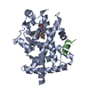
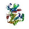
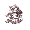
[English] 日本語
Yorodumi
- PDB-6izn: Crystal structure of the PPARgamma-LBD complexed with compound 3g -

+
Open data


ID or keywords:

Loading...

-
Basic information

Entry
Database: PDB / ID: 6izn
TitleCrystal structure of the PPARgamma-LBD complexed with compound 3g
Components
  • Peptide from Peroxisome proliferator-activated receptor gamma coactivator 1-alpha
  • Peroxisome proliferator-activated receptor gamma
KeywordsTRANSCRIPTION
Function / homology
Function and homology information


Regulation of MITF-M dependent genes involved in metabolism / positive regulation of fatty acid oxidation / cellular respiration / prostaglandin receptor activity / negative regulation of receptor signaling pathway via STAT / MECP2 regulates transcription factors / negative regulation of vascular endothelial cell proliferation / negative regulation of extracellular matrix assembly / negative regulation of connective tissue replacement involved in inflammatory response wound healing / Activation of PPARGC1A (PGC-1alpha) by phosphorylation ...Regulation of MITF-M dependent genes involved in metabolism / positive regulation of fatty acid oxidation / cellular respiration / prostaglandin receptor activity / negative regulation of receptor signaling pathway via STAT / MECP2 regulates transcription factors / negative regulation of vascular endothelial cell proliferation / negative regulation of extracellular matrix assembly / negative regulation of connective tissue replacement involved in inflammatory response wound healing / Activation of PPARGC1A (PGC-1alpha) by phosphorylation / positive regulation of cholesterol transport / negative regulation of cellular response to transforming growth factor beta stimulus / : / arachidonate binding / positive regulation of adiponectin secretion / negative regulation of cardiac muscle hypertrophy in response to stress / DNA binding domain binding / lipoprotein transport / positive regulation of vascular associated smooth muscle cell apoptotic process / WW domain binding / STAT family protein binding / positive regulation of fatty acid metabolic process / response to lipid / temperature homeostasis / response to muscle activity / lncRNA binding / negative regulation of SMAD protein signal transduction / negative regulation of type II interferon-mediated signaling pathway / LBD domain binding / negative regulation of cholesterol storage / response to starvation / lipid homeostasis / E-box binding / alpha-actinin binding / intracellular glucose homeostasis / negative regulation of vascular associated smooth muscle cell proliferation / fatty acid oxidation / R-SMAD binding / response to dietary excess / monocyte differentiation / negative regulation of blood vessel endothelial cell migration / white fat cell differentiation / negative regulation of macrophage derived foam cell differentiation / negative regulation of lipid storage / cellular response to low-density lipoprotein particle stimulus / negative regulation of BMP signaling pathway / positive regulation of cholesterol efflux / negative regulation of mitochondrial fission / cell fate commitment / negative regulation of osteoblast differentiation / positive regulation of fat cell differentiation / negative regulation of MAPK cascade / adipose tissue development / BMP signaling pathway / long-chain fatty acid transport / FOXO-mediated transcription of oxidative stress, metabolic and neuronal genes / nuclear retinoid X receptor binding / brown fat cell differentiation / Transcriptional regulation of brown and beige adipocyte differentiation by EBF2 / retinoic acid receptor signaling pathway / cell maturation / energy homeostasis / intracellular receptor signaling pathway / digestion / positive regulation of adipose tissue development / hormone-mediated signaling pathway / epithelial cell differentiation / peroxisome proliferator activated receptor signaling pathway / regulation of cellular response to insulin stimulus / response to nutrient / positive regulation of gluconeogenesis / negative regulation of miRNA transcription / peptide binding / RNA splicing / SUMOylation of transcription cofactors / negative regulation of angiogenesis / positive regulation of DNA-binding transcription factor activity / transcription coregulator binding / positive regulation of apoptotic signaling pathway / Regulation of PTEN gene transcription / nuclear receptor binding / respiratory electron transport chain / gluconeogenesis / fatty acid metabolic process / mitochondrion organization / transcription initiation at RNA polymerase II promoter / transcription coregulator activity / placenta development / SUMOylation of intracellular receptors / negative regulation of smooth muscle cell proliferation / negative regulation of transforming growth factor beta receptor signaling pathway / circadian regulation of gene expression / mRNA transcription by RNA polymerase II / Heme signaling / Transcriptional activation of mitochondrial biogenesis / PPARA activates gene expression / regulation of circadian rhythm / PML body / chromatin DNA binding / Nuclear Receptor transcription pathway
Similarity search - Function
PGC-1alpha, RNA recognition motif / PGC-1 / Peroxisome proliferator-activated receptor gamma / Peroxisome proliferator-activated receptor gamma, N-terminal / PPAR gamma N-terminal region / Peroxisome proliferator-activated receptor / : / Retinoid X Receptor / Retinoid X Receptor / RNA recognition motif ...PGC-1alpha, RNA recognition motif / PGC-1 / Peroxisome proliferator-activated receptor gamma / Peroxisome proliferator-activated receptor gamma, N-terminal / PPAR gamma N-terminal region / Peroxisome proliferator-activated receptor / : / Retinoid X Receptor / Retinoid X Receptor / RNA recognition motif / RNA recognition motif / Eukaryotic RNA Recognition Motif (RRM) profile. / RNA recognition motif domain / RNA-binding domain superfamily / Nuclear hormone receptor / Nuclear hormones receptors DNA-binding region signature. / Zinc finger, nuclear hormone receptor-type / Double treble clef zinc finger, C4 type / Nuclear hormone receptors DNA-binding domain profile. / c4 zinc finger in nuclear hormone receptors / Nuclear hormone receptor, ligand-binding domain / Nuclear hormone receptor-like domain superfamily / Ligand-binding domain of nuclear hormone receptor / Nuclear receptor (NR) ligand-binding (LBD) domain profile. / Ligand binding domain of hormone receptors / Zinc finger, NHR/GATA-type / Nucleotide-binding alpha-beta plait domain superfamily / Orthogonal Bundle / Mainly Alpha
Similarity search - Domain/homology
Chem-B1O / Peroxisome proliferator-activated receptor gamma / Peroxisome proliferator-activated receptor gamma coactivator 1-alpha
Similarity search - Component
Biological speciesHomo sapiens (human)
MethodX-RAY DIFFRACTION / MOLECULAR REPLACEMENT / Resolution: 1.75 Å
AuthorsMatsui, Y. / Hanzawa, H.
CitationJournal: Acs Med.Chem.Lett. / Year: 2019
Title: Structure-Activity Relationship Studies of 3- or 4-Pyridine Derivatives of DS-6930.
Authors: Shinozuka, T. / Tsukada, T. / Fujii, K. / Tokumaru, E. / Matsui, Y. / Wakimoto, S. / Ogata, T. / Araki, K. / Sawamura, R. / Watanabe, N. / Mori, M. / Tanaka, J.
History
DepositionDec 20, 2018Deposition site: PDBJ / Processing site: PDBJ
Revision 1.0Mar 27, 2019Provider: repository / Type: Initial release
Revision 1.1Nov 20, 2019Group: Database references / Category: citation / Item: _citation.pdbx_database_id_PubMed / _citation.title
Revision 1.2Nov 22, 2023Group: Data collection / Database references / Refinement description
Category: chem_comp_atom / chem_comp_bond ...chem_comp_atom / chem_comp_bond / database_2 / pdbx_initial_refinement_model
Item: _database_2.pdbx_DOI / _database_2.pdbx_database_accession

-
Structure visualization

Structure viewerMolecule:
MolmilJmol/JSmol

Downloads & links

-
Assembly

Deposited unit
A: Peroxisome proliferator-activated receptor gamma
C: Peptide from Peroxisome proliferator-activated receptor gamma coactivator 1-alpha
hetero molecules


Theoretical massNumber of molelcules
Total (without water)34,9013
Polymers34,4802
Non-polymers4211
Water2,864159
1


  • Idetical with deposited unit
  • defined by author&software
TypeNameSymmetry operationNumber
identity operation1_555x,y,z1
Buried area1000 Å2
ΔGint-8 kcal/mol
Surface area13520 Å2
MethodPISA
Unit cell
Length a, b, c (Å)38.994, 54.353, 66.594
Angle α, β, γ (deg.)90.00, 92.60, 90.00
Int Tables number4
Space group name H-MP1211

-
Components

#1: Protein Peroxisome proliferator-activated receptor gamma / PPAR-gamma / Nuclear receptor subfamily 1 group C member 3


Mass: 32412.576 Da / Num. of mol.: 1
Source method: isolated from a genetically manipulated source
Source: (gene. exp.) Homo sapiens (human) / Gene: PPARG, NR1C3 / Production host: Escherichia coli (E. coli) / References: UniProt: P37231
#2: Protein/peptide Peptide from Peroxisome proliferator-activated receptor gamma coactivator 1-alpha / PPARGC-1-alpha / Ligand effect modulator 6


Mass: 2067.381 Da / Num. of mol.: 1 / Source method: obtained synthetically / Source: (synth.) Homo sapiens (human) / References: UniProt: Q9UBK2
#3: Chemical ChemComp-B1O / 3-[[6-(2,6-dimethylpyridin-3-yl)oxy-7-fluoranyl-1-methyl-benzimidazol-2-yl]methoxy]benzoic acid


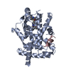
Mass: 421.421 Da / Num. of mol.: 1 / Source method: obtained synthetically / Formula: C23H20FN3O4 / Feature type: SUBJECT OF INVESTIGATION
#4: Water ChemComp-HOH / water


Mass: 18.015 Da / Num. of mol.: 159 / Source method: isolated from a natural source / Formula: H2O

-
Experimental details

-
Experiment

ExperimentMethod: X-RAY DIFFRACTION / Number of used crystals: 1

-
Sample preparation

CrystalDensity Matthews: 2.04 Å3/Da / Density % sol: 39.84 %
Crystal growTemperature: 295 K / Method: vapor diffusion, hanging drop / pH: 8.5 / Details: PEG 4000, NaSCN, Tris-HCl

-
Data collection

DiffractionMean temperature: 93 K / Serial crystal experiment: N
Diffraction sourceSource: ROTATING ANODE / Type: RIGAKU FR-E DW / Wavelength: 1.5418 Å
DetectorType: RIGAKU RAXIS IV / Detector: IMAGE PLATE / Date: Apr 11, 2008
RadiationProtocol: SINGLE WAVELENGTH / Monochromatic (M) / Laue (L): M / Scattering type: x-ray
Radiation wavelengthWavelength: 1.5418 Å / Relative weight: 1
ReflectionResolution: 1.75→50 Å / Num. obs: 27876 / % possible obs: 98 % / Redundancy: 7.5 % / Biso Wilson estimate: 24.3 Å2 / Rsym value: 0.055 / Net I/σ(I): 24.1
Reflection shellResolution: 1.75→1.81 Å / Mean I/σ(I) obs: 3.6 / Num. unique obs: 2625 / Rsym value: 0.259 / % possible all: 93

-
Processing

Software
NameVersionClassification
CNX2005refinement
HKL-2000data reduction
HKL-2000data scaling
CNXphasing
RefinementMethod to determine structure: MOLECULAR REPLACEMENT
Starting model: 3V9T
Resolution: 1.75→19.66 Å / Rfactor Rfree error: 0.005 / Data cutoff high absF: 890501.08 / Data cutoff low absF: 0 / Cross valid method: THROUGHOUT / σ(F): 0
RfactorNum. reflection% reflectionSelection details
Rfree0.265 2785 10.1 %RANDOM
Rwork0.236 ---
obs0.239 27679 98.2 %-
Solvent computationBsol: 52.6396 Å2 / ksol: 0.416923 e/Å3
Displacement parametersBiso mean: 25 Å2
Baniso -1Baniso -2Baniso -3
1-5.46 Å20 Å2-3.55 Å2
2---1.93 Å20 Å2
3----3.53 Å2
Refine analyze
FreeObs
Luzzati coordinate error0.27 Å0.23 Å
Luzzati d res low-5 Å
Luzzati sigma a0.16 Å0.09 Å
Refinement stepCycle: 1 / Resolution: 1.75→19.66 Å
ProteinNucleic acidLigandSolventTotal
Num. atoms2205 0 31 159 2395
Refine LS restraints
Refine-IDTypeDev idealDev ideal target
X-RAY DIFFRACTIONc_bond_d0.004
X-RAY DIFFRACTIONc_bond_d_na
X-RAY DIFFRACTIONc_bond_d_prot
X-RAY DIFFRACTIONc_angle_d
X-RAY DIFFRACTIONc_angle_d_na
X-RAY DIFFRACTIONc_angle_d_prot
X-RAY DIFFRACTIONc_angle_deg0.7
X-RAY DIFFRACTIONc_angle_deg_na
X-RAY DIFFRACTIONc_angle_deg_prot
X-RAY DIFFRACTIONc_dihedral_angle_d18.2
X-RAY DIFFRACTIONc_dihedral_angle_d_na
X-RAY DIFFRACTIONc_dihedral_angle_d_prot
X-RAY DIFFRACTIONc_improper_angle_d0.61
X-RAY DIFFRACTIONc_improper_angle_d_na
X-RAY DIFFRACTIONc_improper_angle_d_prot
X-RAY DIFFRACTIONc_mcbond_it1.11.5
X-RAY DIFFRACTIONc_mcangle_it1.662
X-RAY DIFFRACTIONc_scbond_it1.762
X-RAY DIFFRACTIONc_scangle_it2.532.5
LS refinement shellResolution: 1.75→1.86 Å / Rfactor Rfree error: 0.016 / Total num. of bins used: 6
RfactorNum. reflection% reflection
Rfree0.324 432 9.7 %
Rwork0.294 4026 -
obs--95.4 %
Xplor file
Refine-IDSerial noParam fileTopol file
X-RAY DIFFRACTION1protein_rep.paraprotein.top
X-RAY DIFFRACTION2RTC-3350.prmRTC-3350.top
X-RAY DIFFRACTION3water_rep.paramwater.top

+
About Yorodumi

-
News

-
Feb 9, 2022. New format data for meta-information of EMDB entries

New format data for meta-information of EMDB entries

  • Version 3 of the EMDB header file is now the official format.
  • The previous official version 1.9 will be removed from the archive.

Related info.:EMDB header

External links:wwPDB to switch to version 3 of the EMDB data model

-
Aug 12, 2020. Covid-19 info

Covid-19 info

URL: https://pdbj.org/emnavi/covid19.php

New page: Covid-19 featured information page in EM Navigator.

Related info.:Covid-19 info / Mar 5, 2020. Novel coronavirus structure data

+
Mar 5, 2020. Novel coronavirus structure data

Novel coronavirus structure data

Related info.:Yorodumi Speices / Aug 12, 2020. Covid-19 info

External links:COVID-19 featured content - PDBj / Molecule of the Month (242):Coronavirus Proteases

+
Jan 31, 2019. EMDB accession codes are about to change! (news from PDBe EMDB page)

EMDB accession codes are about to change! (news from PDBe EMDB page)

  • The allocation of 4 digits for EMDB accession codes will soon come to an end. Whilst these codes will remain in use, new EMDB accession codes will include an additional digit and will expand incrementally as the available range of codes is exhausted. The current 4-digit format prefixed with “EMD-” (i.e. EMD-XXXX) will advance to a 5-digit format (i.e. EMD-XXXXX), and so on. It is currently estimated that the 4-digit codes will be depleted around Spring 2019, at which point the 5-digit format will come into force.
  • The EM Navigator/Yorodumi systems omit the EMD- prefix.

Related info.:Q: What is EMD? / ID/Accession-code notation in Yorodumi/EM Navigator

External links:EMDB Accession Codes are Changing Soon! / Contact to PDBj

+
Jul 12, 2017. Major update of PDB

Major update of PDB

  • wwPDB released updated PDB data conforming to the new PDBx/mmCIF dictionary.
  • This is a major update changing the version number from 4 to 5, and with Remediation, in which all the entries are updated.
  • In this update, many items about electron microscopy experimental information are reorganized (e.g. em_software).
  • Now, EM Navigator and Yorodumi are based on the updated data.

External links:wwPDB Remediation / Enriched Model Files Conforming to OneDep Data Standards Now Available in the PDB FTP Archive

-
Yorodumi

Thousand views of thousand structures

  • Yorodumi is a browser for structure data from EMDB, PDB, SASBDB, etc.
  • This page is also the successor to EM Navigator detail page, and also detail information page/front-end page for Omokage search.
  • The word "yorodu" (or yorozu) is an old Japanese word meaning "ten thousand". "mi" (miru) is to see.

Related info.:EMDB / PDB / SASBDB / Comparison of 3 databanks / Yorodumi Search / Aug 31, 2016. New EM Navigator & Yorodumi / Yorodumi Papers / Jmol/JSmol / Function and homology information / Changes in new EM Navigator and Yorodumi

Read more