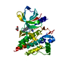
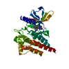
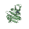
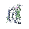
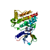
[English] 日本語
Yorodumi
- PDB-5cy3: SYK catalytic domain complexed with a potent and orally bioavaila... -

+
Open data


ID or keywords:

Loading...

-
Basic information

Entry
Database: PDB / ID: 5cy3
TitleSYK catalytic domain complexed with a potent and orally bioavailable benzisothiazole inhibitor
ComponentsTyrosine-protein kinase SYK
KeywordsTransferase/Transferase inhibitor / Transferase-Transferase inhibitor complex
Function / homology
Function and homology information


serotonin secretion by platelet / interleukin-15 receptor binding / positive regulation of interleukin-3 production / regulation of superoxide anion generation / regulation of neutrophil degranulation / regulation of arachidonic acid secretion / cellular response to lectin / gamma-delta T cell differentiation / positive regulation of gamma-delta T cell differentiation / B cell receptor complex ...serotonin secretion by platelet / interleukin-15 receptor binding / positive regulation of interleukin-3 production / regulation of superoxide anion generation / regulation of neutrophil degranulation / regulation of arachidonic acid secretion / cellular response to lectin / gamma-delta T cell differentiation / positive regulation of gamma-delta T cell differentiation / B cell receptor complex / neutrophil activation involved in immune response / Toll-like receptor binding / regulation of platelet aggregation / positive regulation of alpha-beta T cell proliferation / leukocyte activation involved in immune response / positive regulation of mast cell cytokine production / regulation of platelet activation / lymph vessel development / collagen-activated tyrosine kinase receptor signaling pathway / cell activation / positive regulation of mast cell degranulation / macrophage activation involved in immune response / beta selection / regulation of phagocytosis / positive regulation of killing of cells of another organism / leukotriene biosynthetic process / cellular response to molecule of fungal origin / regulation of tumor necrosis factor-mediated signaling pathway / FLT3 signaling through SRC family kinases / early phagosome / interleukin-3-mediated signaling pathway / cellular response to lipid / positive regulation of monocyte chemotactic protein-1 production / regulation of DNA-binding transcription factor activity / positive regulation of cell adhesion mediated by integrin / positive regulation of granulocyte macrophage colony-stimulating factor production / Fc epsilon receptor (FCERI) signaling / Interleukin-2 signaling / positive regulation of alpha-beta T cell differentiation / blood vessel morphogenesis / T cell receptor complex / positive regulation of B cell differentiation / leukocyte cell-cell adhesion / mast cell degranulation / Fc-gamma receptor signaling pathway involved in phagocytosis / Dectin-2 family / Fc-epsilon receptor signaling pathway / stimulatory C-type lectin receptor signaling pathway / phospholipase binding / positive regulation of interleukin-10 production / positive regulation of cysteine-type endopeptidase activity involved in apoptotic process / positive regulation of receptor internalization / cellular response to low-density lipoprotein particle stimulus / FCGR activation / positive regulation of type I interferon production / positive regulation of interleukin-4 production / positive regulation of bone resorption / phosphatase binding / Role of phospholipids in phagocytosis / Role of LAT2/NTAL/LAB on calcium mobilization / positive regulation of calcium-mediated signaling / Signaling by CSF3 (G-CSF) / negative regulation of inflammatory response to antigenic stimulus / GPVI-mediated activation cascade / positive regulation of TORC1 signaling / extrinsic component of cytoplasmic side of plasma membrane / positive regulation of interleukin-12 production / phosphotyrosine residue binding / SH2 domain binding / Integrin signaling / FCERI mediated Ca+2 mobilization / neutrophil chemotaxis / regulation of ERK1 and ERK2 cascade / B cell differentiation / FCGR3A-mediated IL10 synthesis / positive regulation of superoxide anion generation / Antigen activates B Cell Receptor (BCR) leading to generation of second messengers / integrin-mediated signaling pathway / positive regulation of interleukin-8 production / calcium-mediated signaling / Regulation of signaling by CBL / positive regulation of protein-containing complex assembly / FCERI mediated MAPK activation / FCGR3A-mediated phagocytosis / animal organ morphogenesis / B cell receptor signaling pathway / non-specific protein-tyrosine kinase / non-membrane spanning protein tyrosine kinase activity / Inactivation of CSF3 (G-CSF) signaling / receptor internalization / Regulation of actin dynamics for phagocytic cup formation / platelet activation / CLEC7A (Dectin-1) signaling / positive regulation of interleukin-6 production / protein import into nucleus / peptidyl-tyrosine phosphorylation / cell surface receptor protein tyrosine kinase signaling pathway / positive regulation of peptidyl-tyrosine phosphorylation / integrin binding / positive regulation of tumor necrosis factor production
Similarity search - Function
Tyrosine-protein kinase, non-receptor SYK/ZAP-70 / Tyrosine-protein kinase SYK/ZAP-70, inter-SH2 domain superfamily / SYK/ZAP-70, N-terminal SH2 domain / SH2 domain / Src homology 2 (SH2) domain profile. / Src homology 2 domains / SH2 domain / SH2 domain superfamily / Tyrosine-protein kinase, catalytic domain / Tyrosine kinase, catalytic domain ...Tyrosine-protein kinase, non-receptor SYK/ZAP-70 / Tyrosine-protein kinase SYK/ZAP-70, inter-SH2 domain superfamily / SYK/ZAP-70, N-terminal SH2 domain / SH2 domain / Src homology 2 (SH2) domain profile. / Src homology 2 domains / SH2 domain / SH2 domain superfamily / Tyrosine-protein kinase, catalytic domain / Tyrosine kinase, catalytic domain / Tyrosine protein kinases specific active-site signature. / Tyrosine-protein kinase, active site / Protein tyrosine and serine/threonine kinase / Serine-threonine/tyrosine-protein kinase, catalytic domain / Transferase(Phosphotransferase) domain 1 / Transferase(Phosphotransferase); domain 1 / Phosphorylase Kinase; domain 1 / Phosphorylase Kinase; domain 1 / Protein kinase, ATP binding site / Protein kinases ATP-binding region signature. / Protein kinase domain profile. / Protein kinase domain / Protein kinase-like domain superfamily / 2-Layer Sandwich / Orthogonal Bundle / Mainly Alpha / Alpha Beta
Similarity search - Domain/homology
Chem-55Y / Tyrosine-protein kinase SYK
Similarity search - Component
Biological speciesHomo sapiens (human)
MethodX-RAY DIFFRACTION / SYNCHROTRON / MOLECULAR REPLACEMENT / Resolution: 1.76 Å
AuthorsLee, C.C.
CitationJournal: Bioorg.Med.Chem.Lett. / Year: 2015
Title: Orally bioavailable Syk inhibitors with activity in a rat PK/PD model.
Authors: Thoma, G. / Veenstra, S. / Strang, R. / Blanz, J. / Vangrevelinghe, E. / Berghausen, J. / Lee, C.C. / Zerwes, H.G.
History
DepositionJul 30, 2015Deposition site: RCSB / Processing site: RCSB
Revision 1.0Sep 23, 2015Provider: repository / Type: Initial release
Revision 1.1Oct 28, 2015Group: Database references
Revision 1.2Dec 16, 2015Group: Refinement description
Revision 1.3Mar 6, 2024Group: Data collection / Database references / Derived calculations
Category: chem_comp_atom / chem_comp_bond ...chem_comp_atom / chem_comp_bond / database_2 / pdbx_struct_oper_list
Item: _database_2.pdbx_DOI / _database_2.pdbx_database_accession / _pdbx_struct_oper_list.symmetry_operation

-
Structure visualization

Structure viewerMolecule:
MolmilJmol/JSmol

Downloads & links

-
Assembly

Deposited unit
A: Tyrosine-protein kinase SYK
hetero molecules


Theoretical massNumber of molelcules
Total (without water)34,2002
Polymers33,8561
Non-polymers3441
Water3,477193
1


  • Idetical with deposited unit
  • defined by author
TypeNameSymmetry operationNumber
identity operation1_555x,y,z1
Unit cell
Length a, b, c (Å)41.504, 37.656, 90.888
Angle α, β, γ (deg.)90.000, 100.140, 90.000
Int Tables number4
Space group name H-MP1211

-
Components

#1: Protein Tyrosine-protein kinase SYK / / Spleen tyrosine kinase / p72-Syk


Mass: 33855.930 Da / Num. of mol.: 1 / Fragment: residues 356-635 / Mutation: K467T
Source method: isolated from a genetically manipulated source
Source: (gene. exp.) Homo sapiens (human) / Gene: SYK / Plasmid: pFastBac1 / Cell line (production host): Sf9 / Production host: 
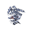
Spodoptera frugiperda (fall armyworm)
References: UniProt: P43405, non-specific protein-tyrosine kinase
#2: Chemical ChemComp-55Y / (5R)-5-[(1R)-1-{[6-(1-methyl-1H-pyrazol-4-yl)-2,1-benzothiazol-4-yl]oxy}ethyl]-1,3-oxazolidin-2-one


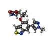
Mass: 344.388 Da / Num. of mol.: 1 / Source method: obtained synthetically / Formula: C16H16N4O3S
#3: Water ChemComp-HOH / water / Water


Mass: 18.015 Da / Num. of mol.: 193 / Source method: isolated from a natural source / Formula: H2O

-
Experimental details

-
Experiment

ExperimentMethod: X-RAY DIFFRACTION / Number of used crystals: 1

-
Sample preparation

CrystalDensity Matthews: 2.17 Å3/Da / Density % sol: 43.38 %
Crystal growTemperature: 293 K / Method: vapor diffusion, sitting drop / pH: 7.1 / Details: PEG 3350 20%, ammonium acetate 0.2M

-
Data collection

DiffractionMean temperature: 100 K
Diffraction sourceSource: SYNCHROTRON / Site: ALS / Beamline: 5.0.3 / Wavelength: 0.9765 Å
DetectorType: ADSC QUANTUM 315r / Detector: CCD / Date: Apr 17, 2014
RadiationProtocol: SINGLE WAVELENGTH / Monochromatic (M) / Laue (L): M / Scattering type: x-ray
Radiation wavelengthWavelength: 0.9765 Å / Relative weight: 1
ReflectionResolution: 1.75→50 Å / Num. obs: 27534 / % possible obs: 98.9 % / Redundancy: 2.6 % / Biso Wilson estimate: 21.27 Å2 / Rmerge(I) obs: 0.041 / Rpim(I) all: 0.031 / Rrim(I) all: 0.052 / Χ2: 0.932 / Net I/av σ(I): 22.387 / Net I/σ(I): 12.4 / Num. measured all: 72734
Reflection shell

Diffraction-ID: 1 / Rejects: 0

Resolution (Å)Redundancy (%)Rmerge(I) obsNum. unique allCC1/2Rpim(I) allRrim(I) allΧ2% possible all
1.75-1.792.50.41418110.80.3150.5230.95198.3
1.79-1.842.70.31317890.8770.2290.390.95798.1
1.84-1.892.70.25218190.9110.1850.314198.3
1.89-1.942.70.19517670.9480.1450.2441.02498.4
1.94-22.70.15118410.9650.110.1871.0198.8
2-2.072.60.11318490.9740.0840.1411.01599.1
2.07-2.162.70.08918110.9860.0650.1110.96199
2.16-2.262.70.07218230.9890.0530.090.92899.2
2.26-2.382.70.06118400.9920.0450.0760.86399.4
2.38-2.522.60.0518490.9920.0370.0630.98199.6
2.52-2.722.70.0418440.9950.0290.050.84799.9
2.72-2.992.60.03618520.9960.0270.0460.89499.7
2.99-3.432.60.03718850.9960.0270.0460.90199.6
3.43-4.322.60.03318500.9960.0250.0420.87699
4.32-502.60.02619040.9970.020.0330.78697.4

-
Processing

Software
NameVersionClassification
DENZOdata reduction
SCALEPACKdata scaling
BUSTER-TNT2.11.5refinement
PDB_EXTRACT3.15data extraction
HKL-2000data reduction
REFMACphasing
RefinementMethod to determine structure: MOLECULAR REPLACEMENT / Resolution: 1.76→39.91 Å / Cor.coef. Fo:Fc: 0.9559 / Cor.coef. Fo:Fc free: 0.947 / SU R Cruickshank DPI: 0.113 / Cross valid method: THROUGHOUT / σ(F): 0 / SU R Blow DPI: 0.117 / SU Rfree Blow DPI: 0.105 / SU Rfree Cruickshank DPI: 0.104
RfactorNum. reflection% reflectionSelection details
Rfree0.1947 1319 4.93 %RANDOM
Rwork0.1699 ---
obs0.1711 26766 96.32 %-
Displacement parametersBiso max: 119.61 Å2 / Biso mean: 26.13 Å2 / Biso min: 8.1 Å2
Baniso -1Baniso -2Baniso -3
1--1.1176 Å20 Å20.6773 Å2
2--1.5699 Å20 Å2
3----0.4523 Å2
Refine analyzeLuzzati coordinate error obs: 0.193 Å
Refinement stepCycle: final / Resolution: 1.76→39.91 Å
ProteinNucleic acidLigandSolventTotal
Num. atoms2185 0 24 193 2402
Biso mean--16.36 34.28 -
Num. residues----276
Refine LS restraints
Refine-IDTypeNumberRestraint functionWeightDev ideal
X-RAY DIFFRACTIONt_dihedral_angle_d784SINUSOIDAL2
X-RAY DIFFRACTIONt_trig_c_planes54HARMONIC2
X-RAY DIFFRACTIONt_gen_planes348HARMONIC5
X-RAY DIFFRACTIONt_it2277HARMONIC20
X-RAY DIFFRACTIONt_nbd
X-RAY DIFFRACTIONt_improper_torsion
X-RAY DIFFRACTIONt_pseud_angle
X-RAY DIFFRACTIONt_chiral_improper_torsion276SEMIHARMONIC5
X-RAY DIFFRACTIONt_sum_occupancies3HARMONIC1
X-RAY DIFFRACTIONt_utility_distance
X-RAY DIFFRACTIONt_utility_angle
X-RAY DIFFRACTIONt_utility_torsion
X-RAY DIFFRACTIONt_ideal_dist_contact2689SEMIHARMONIC4
X-RAY DIFFRACTIONt_bond_d2277HARMONIC20.01
X-RAY DIFFRACTIONt_angle_deg3085HARMONIC20.95
X-RAY DIFFRACTIONt_omega_torsion3.02
X-RAY DIFFRACTIONt_other_torsion15.49
LS refinement shellResolution: 1.76→1.83 Å / Total num. of bins used: 13
RfactorNum. reflection% reflection
Rfree0.2273 106 4.41 %
Rwork0.194 2296 -
all0.1954 2402 -
obs--96.32 %
Refinement TLS params.Method: refined / Details: Chain A catalytic domain / Origin x: 1.2456 Å / Origin y: -7.6394 Å / Origin z: -24.0533 Å
111213212223313233
T-0.0836 Å20.0073 Å20.0005 Å2--0.0699 Å20.0131 Å2---0.0661 Å2
L1.347 °20.2662 °20.1474 °2-0.926 °20.1102 °2--1.5144 °2
S0.063 Å °-0.0162 Å °-0.0228 Å °0.1368 Å °-0.0493 Å °0.0234 Å °-0.0276 Å °-0.1562 Å °-0.0137 Å °
Refinement TLS groupSelection details: { A|360 - A|637 }

+
About Yorodumi

-
News

-
Feb 9, 2022. New format data for meta-information of EMDB entries

New format data for meta-information of EMDB entries

  • Version 3 of the EMDB header file is now the official format.
  • The previous official version 1.9 will be removed from the archive.

Related info.:EMDB header

External links:wwPDB to switch to version 3 of the EMDB data model

-
Aug 12, 2020. Covid-19 info

Covid-19 info

URL: https://pdbj.org/emnavi/covid19.php

New page: Covid-19 featured information page in EM Navigator.

Related info.:Covid-19 info / Mar 5, 2020. Novel coronavirus structure data

+
Mar 5, 2020. Novel coronavirus structure data

Novel coronavirus structure data

Related info.:Yorodumi Speices / Aug 12, 2020. Covid-19 info

External links:COVID-19 featured content - PDBj / Molecule of the Month (242):Coronavirus Proteases

+
Jan 31, 2019. EMDB accession codes are about to change! (news from PDBe EMDB page)

EMDB accession codes are about to change! (news from PDBe EMDB page)

  • The allocation of 4 digits for EMDB accession codes will soon come to an end. Whilst these codes will remain in use, new EMDB accession codes will include an additional digit and will expand incrementally as the available range of codes is exhausted. The current 4-digit format prefixed with “EMD-” (i.e. EMD-XXXX) will advance to a 5-digit format (i.e. EMD-XXXXX), and so on. It is currently estimated that the 4-digit codes will be depleted around Spring 2019, at which point the 5-digit format will come into force.
  • The EM Navigator/Yorodumi systems omit the EMD- prefix.

Related info.:Q: What is EMD? / ID/Accession-code notation in Yorodumi/EM Navigator

External links:EMDB Accession Codes are Changing Soon! / Contact to PDBj

+
Jul 12, 2017. Major update of PDB

Major update of PDB

  • wwPDB released updated PDB data conforming to the new PDBx/mmCIF dictionary.
  • This is a major update changing the version number from 4 to 5, and with Remediation, in which all the entries are updated.
  • In this update, many items about electron microscopy experimental information are reorganized (e.g. em_software).
  • Now, EM Navigator and Yorodumi are based on the updated data.

External links:wwPDB Remediation / Enriched Model Files Conforming to OneDep Data Standards Now Available in the PDB FTP Archive

-
Yorodumi

Thousand views of thousand structures

  • Yorodumi is a browser for structure data from EMDB, PDB, SASBDB, etc.
  • This page is also the successor to EM Navigator detail page, and also detail information page/front-end page for Omokage search.
  • The word "yorodu" (or yorozu) is an old Japanese word meaning "ten thousand". "mi" (miru) is to see.

Related info.:EMDB / PDB / SASBDB / Comparison of 3 databanks / Yorodumi Search / Aug 31, 2016. New EM Navigator & Yorodumi / Yorodumi Papers / Jmol/JSmol / Function and homology information / Changes in new EM Navigator and Yorodumi

Read more