 Movie
Movie Controller

[English] 日本語
 Yorodumi
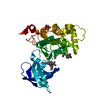
- PDB-4zck: Crystal Structure of C-terminal Fragment of Escherichia coli BipA/TypA -
+ Open data

- Basic information
| Entry | Database: PDB / ID: 4zck | ||||||
|---|---|---|---|---|---|---|---|
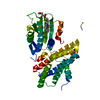
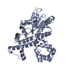
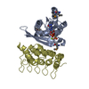
| Title | Crystal Structure of C-terminal Fragment of Escherichia coli BipA/TypA | ||||||
 Components Components | GTP-binding protein TypA/BipA | ||||||
 Keywords Keywords | GTP-BINDING PROTEIN / BipA / GTPase / Nucleotide | ||||||
| Function / homology |  Function and homology information Function and homology informationguanosine tetraphosphate binding / response to cold / protein folding chaperone / response to heat / ribosome binding / ribosomal large subunit assembly / Hydrolases; Acting on acid anhydrides; Acting on GTP to facilitate cellular and subcellular movement / tRNA binding / rRNA binding / translation ...guanosine tetraphosphate binding / response to cold / protein folding chaperone / response to heat / ribosome binding / ribosomal large subunit assembly / Hydrolases; Acting on acid anhydrides; Acting on GTP to facilitate cellular and subcellular movement / tRNA binding / rRNA binding / translation / ribonucleoprotein complex / GTPase activity / GTP binding / cytosol / cytoplasm Similarity search - Function | ||||||
| Biological species |  | ||||||
| Method |  X-RAY DIFFRACTION / SYNCHROTRON / MOLECULAR REPLACEMENT / Resolution: 2.48 Å X-RAY DIFFRACTION / SYNCHROTRON / MOLECULAR REPLACEMENT / Resolution: 2.48 Å | ||||||
 Authors Authors | Fan, H.T. / Hahm, J. / Diggs, S. / Blaha, G. | ||||||
 Citation Citation | Journal: J.Biol.Chem. / Year: 2015 Title: Structural and Functional Analysis of BipA, a Regulator of Virulence in Enteropathogenic Escherichia coli. Authors: Fan, H. / Hahm, J. / Diggs, S. / Perry, J.J. / Blaha, G. | ||||||
| History |
|
- Structure visualization
| Structure viewer | Molecule: MolmilJmol/JSmol |
|---|
- Downloads & links
-Download
| PDBx/mmCIF format | 4zck.cif.gz | 137.1 KB | Display | PDBx/mmCIF format |
|---|---|---|---|---|
| PDB format | pdb4zck.ent.gz | 105.5 KB | Display | PDB format |
| PDBx/mmJSON format | 4zck.json.gz | Tree view | PDBx/mmJSON format | |
| Others |  Other downloads Other downloads |
-Validation report
| Arichive directory | https://data.pdbj.org/pub/pdb/validation_reports/zc/4zckftp://data.pdbj.org/pub/pdb/validation_reports/zc/4zck | HTTPS FTP |
|---|
-Related structure data
| Related structure data |  4zciC  4zclC 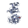 4zcmC  3e3xS C: citing same article ( S: Starting model for refinement |
|---|---|
| Similar structure data |


-Links
 PDBj
PDBj


- Assembly
| Deposited unit | 
| ||||||||
|---|---|---|---|---|---|---|---|---|---|
| 1 |
| ||||||||
| Unit cell |
|
-Components
| #1: Protein | Mass: 37040.930 Da / Num. of mol.: 1 / Fragment: UNP residues 306-603 Source method: isolated from a genetically manipulated source Source: (gene. exp.) Strain: K12 / Gene: typA, bipA, yihK, b3871, JW5571 / Production host: | ||
|---|---|---|---|
| #2: Chemical |   Mass: 24.305 Da / Num. of mol.: 2 Mass: 24.305 Da / Num. of mol.: 2Source method: isolated from a genetically manipulated source Formula: Mg #3: Water | ChemComp-HOH / |  Mass: 18.015 Da / Num. of mol.: 33 / Source method: isolated from a natural source / Formula: H2O Mass: 18.015 Da / Num. of mol.: 33 / Source method: isolated from a natural source / Formula: H2O |
-Experimental details
-Experiment
| Experiment | Method: X-RAY DIFFRACTION / Number of used crystals: 1 |
|---|
- Sample preparation
| Crystal | Density Matthews: 4.52 Å3/Da / Density % sol: 72.78 % |
|---|---|
| Crystal grow | Temperature: 293 K / Method: vapor diffusion, sitting drop / pH: 7.6 Details: 3.04 M sodium formate, 200 mM Tris-HCl, and 1 mM magnesium bromide, pH 7.6 |
-Data collection
| Diffraction | Mean temperature: 100 K |
|---|---|
| Diffraction source | Source: SYNCHROTRON / Site: APS  / Beamline: 24-ID-C / Wavelength: 0.9792 Å / Beamline: 24-ID-C / Wavelength: 0.9792 Å |
| Detector | Type: DECTRIS PILATUS 6M-F / Detector: PIXEL / Date: Feb 23, 2015 |
| Radiation | Monochromator: Si(111) / Protocol: SINGLE WAVELENGTH / Monochromatic (M) / Laue (L): M / Scattering type: x-ray |
| Radiation wavelength | Wavelength: 0.9792 Å / Relative weight: 1 |
| Reflection | Resolution: 2.48→48 Å / Num. obs: 24915 / % possible obs: 99.8 % / Redundancy: 7.5 % / Biso Wilson estimate: 70.4 Å2 / Rmerge(I) obs: 0.14 / Net I/σ(I): 6.5 |
| Reflection shell | Resolution: 2.48→2.58 Å / Redundancy: 5.4 % / Rmerge(I) obs: 1.3 / Mean I/σ(I) obs: 1 / % possible all: 98.7 |
- Processing
| Software |
| |||||||||||||||||||||||||||||||||||||||||||||||||||||||||||||||||||||||||||||||||||||||||||||||||||||||||||||||||||||||||||||||||||||||||||||||||||||||||||||||||||||||||||||||
|---|---|---|---|---|---|---|---|---|---|---|---|---|---|---|---|---|---|---|---|---|---|---|---|---|---|---|---|---|---|---|---|---|---|---|---|---|---|---|---|---|---|---|---|---|---|---|---|---|---|---|---|---|---|---|---|---|---|---|---|---|---|---|---|---|---|---|---|---|---|---|---|---|---|---|---|---|---|---|---|---|---|---|---|---|---|---|---|---|---|---|---|---|---|---|---|---|---|---|---|---|---|---|---|---|---|---|---|---|---|---|---|---|---|---|---|---|---|---|---|---|---|---|---|---|---|---|---|---|---|---|---|---|---|---|---|---|---|---|---|---|---|---|---|---|---|---|---|---|---|---|---|---|---|---|---|---|---|---|---|---|---|---|---|---|---|---|---|---|---|---|---|---|---|---|---|---|
| Refinement | Method to determine structure: MOLECULAR REPLACEMENT Starting model: 3E3X Resolution: 2.48→48 Å / SU ML: 0.38 / Cross valid method: FREE R-VALUE / σ(F): 1.35 / Phase error: 29.04 / Stereochemistry target values: ML
| |||||||||||||||||||||||||||||||||||||||||||||||||||||||||||||||||||||||||||||||||||||||||||||||||||||||||||||||||||||||||||||||||||||||||||||||||||||||||||||||||||||||||||||||
| Solvent computation | Shrinkage radii: 0.9 Å / VDW probe radii: 1.11 Å / Solvent model: FLAT BULK SOLVENT MODEL | |||||||||||||||||||||||||||||||||||||||||||||||||||||||||||||||||||||||||||||||||||||||||||||||||||||||||||||||||||||||||||||||||||||||||||||||||||||||||||||||||||||||||||||||
| Displacement parameters | Biso mean: 86 Å2 | |||||||||||||||||||||||||||||||||||||||||||||||||||||||||||||||||||||||||||||||||||||||||||||||||||||||||||||||||||||||||||||||||||||||||||||||||||||||||||||||||||||||||||||||
| Refinement step | Cycle: LAST / Resolution: 2.48→48 Å
| |||||||||||||||||||||||||||||||||||||||||||||||||||||||||||||||||||||||||||||||||||||||||||||||||||||||||||||||||||||||||||||||||||||||||||||||||||||||||||||||||||||||||||||||
| Refine LS restraints |
| |||||||||||||||||||||||||||||||||||||||||||||||||||||||||||||||||||||||||||||||||||||||||||||||||||||||||||||||||||||||||||||||||||||||||||||||||||||||||||||||||||||||||||||||
| LS refinement shell |
| |||||||||||||||||||||||||||||||||||||||||||||||||||||||||||||||||||||||||||||||||||||||||||||||||||||||||||||||||||||||||||||||||||||||||||||||||||||||||||||||||||||||||||||||
| Refinement TLS params. | Method: refined / Refine-ID: X-RAY DIFFRACTION
| |||||||||||||||||||||||||||||||||||||||||||||||||||||||||||||||||||||||||||||||||||||||||||||||||||||||||||||||||||||||||||||||||||||||||||||||||||||||||||||||||||||||||||||||
| Refinement TLS group |
|
