 Movie
Movie Controller

[English] 日本語
 Yorodumi
- PDB-3e3q: Structure of the 3alpham13 high-affinity mutant of the 2C TCR in ... -
+ Open data

- Basic information
| Entry | Database: PDB / ID: 3e3q | ||||||
|---|---|---|---|---|---|---|---|
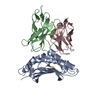
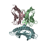
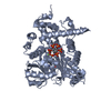
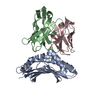
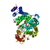
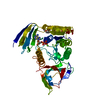
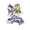
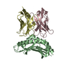
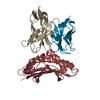
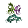
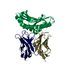
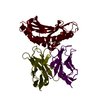
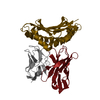
| Title | Structure of the 3alpham13 high-affinity mutant of the 2C TCR in complex with Ld/QL9 | ||||||
 Components Components |
| ||||||
 Keywords Keywords | IMMUNE SYSTEM / TCR / MHC / high affinity / cross reactivity / Glycoprotein / Immune response / Membrane / MHC I / Phosphoprotein / Transmembrane / Immunoglobulin domain / Receptor | ||||||
| Function / homology |  Function and homology information Function and homology informationMHC class Ib protein complex / natural killer cell lectin-like receptor binding / TAP2 binding / TAP1 binding / cis-Golgi network membrane / T cell receptor complex / TAP complex binding / Golgi medial cisterna / CD8 receptor binding / beta-2-microglobulin binding ...MHC class Ib protein complex / natural killer cell lectin-like receptor binding / TAP2 binding / TAP1 binding / cis-Golgi network membrane / T cell receptor complex / TAP complex binding / Golgi medial cisterna / CD8 receptor binding / beta-2-microglobulin binding / endoplasmic reticulum exit site / TAP binding / MHC class I protein binding / antigen processing and presentation of endogenous peptide antigen via MHC class Ib / antigen processing and presentation of endogenous peptide antigen via MHC class I via ER pathway, TAP-independent / T cell receptor binding / 14-3-3 protein binding / lumenal side of endoplasmic reticulum membrane / defense response / MHC class I peptide loading complex / MHC class I protein complex / positive regulation of T cell mediated cytotoxicity / peptide antigen binding / phagocytic vesicle membrane / protein-folding chaperone binding / early endosome membrane / adaptive immune response / early endosome / immune response / receptor ligand activity / signaling receptor binding / Golgi membrane / external side of plasma membrane / lysosomal membrane / cell surface / endoplasmic reticulum / Golgi apparatus / protein homodimerization activity / : / identical protein binding / plasma membrane Similarity search - Function | ||||||
| Biological species |  | ||||||
| Method |  X-RAY DIFFRACTION / SYNCHROTRON / MOLECULAR REPLACEMENT / molecular replacement / Resolution: 2.95 Å X-RAY DIFFRACTION / SYNCHROTRON / MOLECULAR REPLACEMENT / molecular replacement / Resolution: 2.95 Å | ||||||
 Authors Authors | Colf, L.A. / Garcia, K.C. | ||||||
 Citation Citation | Journal: J.Immunol. / Year: 2008 Title: Distinct CDR3 conformations in TCRs determine the level of cross-reactivity for diverse antigens, but not the docking orientation. Authors: Jones, L.L. / Colf, L.A. / Stone, J.D. / Garcia, K.C. / Kranz, D.M. | ||||||
| History |
|
- Structure visualization
| Structure viewer | Molecule: MolmilJmol/JSmol |
|---|
- Downloads & links
-Download
| PDBx/mmCIF format | 3e3q.cif.gz | 620.6 KB | Display | PDBx/mmCIF format |
|---|---|---|---|---|
| PDB format | pdb3e3q.ent.gz | 514.3 KB | Display | PDB format |
| PDBx/mmJSON format | 3e3q.json.gz | Tree view | PDBx/mmJSON format | |
| Others |  Other downloads Other downloads |
-Validation report
| Arichive directory | https://data.pdbj.org/pub/pdb/validation_reports/e3/3e3qftp://data.pdbj.org/pub/pdb/validation_reports/e3/3e3q | HTTPS FTP |
|---|
-Related structure data
| Related structure data |  3e2hC  2oi9S S: Starting model for refinement C: citing same article ( |
|---|---|
| Similar structure data |


-Links
 PDBj
PDBj

- Assembly

-Components
| #1: Protein | Mass: 20541.621 Da / Num. of mol.: 8 / Fragment: UNP residues 25-199 Mutation: F8Y, V12T, P15R, I23T, N30D, A49V, I66V, W97R, K131R Source method: isolated from a genetically manipulated source Source: (gene. exp.)  #2: Protein/peptide | Mass: 1063.202 Da / Num. of mol.: 8 / Source method: obtained synthetically / Details: This sequence occurs naturally in mouse #3: Protein | Mass: 12071.595 Da / Num. of mol.: 8 / Mutation: L43P, W82R, G99D F100P A101P S102P A103L Source method: isolated from a genetically manipulated source Source: (gene. exp.) #4: Protein | Mass: 12039.129 Da / Num. of mol.: 8 / Mutation: G17E, G42E, H47Y, I77T, L81S Source method: isolated from a genetically manipulated source Source: (gene. exp.) Has protein modification | Y | |
|---|
-Experimental details
-Experiment
| Experiment | Method: X-RAY DIFFRACTION / Number of used crystals: 1 |
|---|
- Sample preparation
| Crystal | Density Matthews: 3.1 Å3/Da / Density % sol: 60.37 % |
|---|---|
| Crystal grow | Temperature: 295 K / Method: vapor diffusion, sitting drop Details: 0.2M ammonium citrate, 20% PEG 3350, 3% trimethyl amine N-oxide dihydrate, VAPOR DIFFUSION, SITTING DROP, temperature 295K |
-Data collection
| Diffraction | Mean temperature: 100 K |
|---|---|
| Diffraction source | Source: SYNCHROTRON / Site: SSRL  / Beamline: BL11-1 / Wavelength: 1 Å / Beamline: BL11-1 / Wavelength: 1 Å |
| Detector | Type: MARMOSAIC 325 mm CCD / Detector: CCD / Date: Jan 30, 2007 |
| Radiation | Protocol: SINGLE WAVELENGTH / Monochromatic (M) / Laue (L): M / Scattering type: x-ray |
| Radiation wavelength | Wavelength: 1 Å / Relative weight: 1 |
| Reflection | Resolution: 2.95→50 Å / Num. obs: 93313 / % possible obs: 97.4 % / Redundancy: 4.1 % / Rmerge(I) obs: 0.13 / Χ2: 1.117 / Net I/σ(I): 10.3 |
| Reflection shell | Resolution: 2.95→3.06 Å / Redundancy: 4.1 % / Rmerge(I) obs: 0.656 / Mean I/σ(I) obs: 2 / Num. unique all: 9386 / Χ2: 1.005 / % possible all: 97.3 |
-Phasing
| Phasing | Method: molecular replacement |
|---|
- Processing
| Software |
| ||||||||||||||||||||||||||||||||
|---|---|---|---|---|---|---|---|---|---|---|---|---|---|---|---|---|---|---|---|---|---|---|---|---|---|---|---|---|---|---|---|---|---|
| Refinement | Method to determine structure: MOLECULAR REPLACEMENT Starting model: PDB entry 2OI9 Resolution: 2.95→41.52 Å / Occupancy max: 1 / Occupancy min: 1 / σ(F): 0
| ||||||||||||||||||||||||||||||||
| Solvent computation | Bsol: 23.072 Å2 | ||||||||||||||||||||||||||||||||
| Displacement parameters | Biso max: 100.58 Å2 / Biso mean: 47.947 Å2 / Biso min: 18.91 Å2
| ||||||||||||||||||||||||||||||||
| Refinement step | Cycle: LAST / Resolution: 2.95→41.52 Å
| ||||||||||||||||||||||||||||||||
| Refine LS restraints |
| ||||||||||||||||||||||||||||||||
| Xplor file |
|
