 Movie
Movie Controller


+ Open data

- Basic information
| Entry | Database: PDB / ID: 4ohx | ||||||
|---|---|---|---|---|---|---|---|
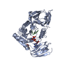
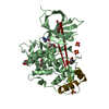
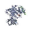
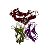
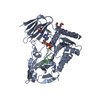
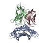
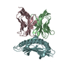
| Title | C. Elegans Clp1 bound to ADP and Mg2+ (RNA released state) | ||||||
 Components Components | Protein clpf-1 | ||||||
 Keywords Keywords | RNA BINDING PROTEIN / polynucleotide kinase | ||||||
| Function / homology |  Function and homology information Function and homology informationProcessing of Capped Intron-Containing Pre-mRNA / Processing of Intronless Pre-mRNAs / mRNA 3'-end processing / RNA Polymerase II Transcription Termination / ATP-dependent polyribonucleotide 5'-hydroxyl-kinase activity / polynucleotide 5'-hydroxyl-kinase activity / mRNA cleavage factor complex / tRNA splicing, via endonucleolytic cleavage and ligation / mRNA 3'-end processing / ATP binding / nucleus Similarity search - Function | ||||||
| Biological species |  | ||||||
| Method |  X-RAY DIFFRACTION / SYNCHROTRON / FOURIER SYNTHESIS / Resolution: 1.98 Å X-RAY DIFFRACTION / SYNCHROTRON / FOURIER SYNTHESIS / Resolution: 1.98 Å | ||||||
 Authors Authors | Dikfidan, A. / Loll, B. / Zeymer, C. / Clausen, T. / Meinhart, A. | ||||||
 Citation Citation | Journal: Mol.Cell / Year: 2014 Title: RNA specificity and regulation of catalysis in the eukaryotic polynucleotide kinase clp1. Authors: Dikfidan, A. / Loll, B. / Zeymer, C. / Magler, I. / Clausen, T. / Meinhart, A. | ||||||
| History |
|
- Structure visualization
| Structure viewer | Molecule: MolmilJmol/JSmol |
|---|
- Downloads & links
-Download
| PDBx/mmCIF format | 4ohx.cif.gz | 179 KB | Display | PDBx/mmCIF format |
|---|---|---|---|---|
| PDB format | pdb4ohx.ent.gz | 140.4 KB | Display | PDB format |
| PDBx/mmJSON format | 4ohx.json.gz | Tree view | PDBx/mmJSON format | |
| Others |  Other downloads Other downloads |
-Validation report
| Arichive directory | https://data.pdbj.org/pub/pdb/validation_reports/oh/4ohxftp://data.pdbj.org/pub/pdb/validation_reports/oh/4ohx | HTTPS FTP |
|---|
-Related structure data
| Related structure data |  4ohvC 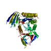 4ohwC  4ohyC  4ohzC  4oi0C  4oi1C  4oi2C  4oi4C C: citing same article ( |
|---|---|
| Similar structure data |


-Links
 PDBj
PDBj- Assembly
| Deposited unit | 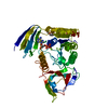
| ||||||||
|---|---|---|---|---|---|---|---|---|---|
| 1 |
| ||||||||
| Unit cell |
|
-Components
| #1: Protein | Mass: 47936.453 Da / Num. of mol.: 1 Source method: isolated from a genetically manipulated source Source: (gene. exp.)  |
|---|---|
| #2: Chemical | ChemComp-ADP /   Mass: 427.201 Da / Num. of mol.: 1 / Source method: obtained synthetically / Formula: C10H15N5O10P2 / Comment: ADP, energy-carrying molecule*YM Mass: 427.201 Da / Num. of mol.: 1 / Source method: obtained synthetically / Formula: C10H15N5O10P2 / Comment: ADP, energy-carrying molecule*YM |
| #3: Chemical | ChemComp-MG /   Mass: 24.305 Da / Num. of mol.: 1 / Source method: obtained synthetically / Formula: Mg Mass: 24.305 Da / Num. of mol.: 1 / Source method: obtained synthetically / Formula: Mg |
| #4: Chemical | ChemComp-2PE /   Mass: 414.488 Da / Num. of mol.: 1 / Source method: obtained synthetically / Formula: C18H38O10 / Comment: precipitant*YM Mass: 414.488 Da / Num. of mol.: 1 / Source method: obtained synthetically / Formula: C18H38O10 / Comment: precipitant*YM |
| #5: Water | ChemComp-HOH /  Mass: 18.015 Da / Num. of mol.: 179 / Source method: isolated from a natural source / Formula: H2O Mass: 18.015 Da / Num. of mol.: 179 / Source method: isolated from a natural source / Formula: H2O |
-Experimental details
-Experiment
| Experiment | Method: X-RAY DIFFRACTION / Number of used crystals: 1 |
|---|
- Sample preparation
| Crystal | Density Matthews: 2.52 Å3/Da / Density % sol: 51.23 % |
|---|---|
| Crystal grow | Temperature: 293 K / Method: vapor diffusion, hanging drop / pH: 5.5 Details: 100 mM Na2HPO4/KH2PO4 200 mM NaCl 15 mM MgCl2 90 mM sarcosine 25% (w/v) PEG 1000, pH 5.5, VAPOR DIFFUSION, HANGING DROP, temperature 293K |
-Data collection
| Diffraction | Mean temperature: 100 K |
|---|---|
| Diffraction source | Source: SYNCHROTRON / Site: SLS  / Beamline: X10SA / Wavelength: 0.9792 Å / Beamline: X10SA / Wavelength: 0.9792 Å |
| Detector | Type: DECTRIS PILATUS 6M / Detector: PIXEL / Date: Jul 30, 2010 |
| Radiation | Monochromator: SI(111) / Protocol: SINGLE WAVELENGTH / Monochromatic (M) / Laue (L): M / Scattering type: x-ray |
| Radiation wavelength | Wavelength: 0.9792 Å / Relative weight: 1 |
| Reflection | Resolution: 1.98→50 Å / Num. obs: 30056 / % possible obs: 98.6 % / Observed criterion σ(I): -3 / Redundancy: 3.5 % / Biso Wilson estimate: 45.8 Å2 |
| Reflection shell | Resolution: 2→2.1 Å / Redundancy: 3.5 % / % possible all: 99.1 |
- Processing
| Software |
| ||||||||||||||||||||||||||||||||||||||||||||||||||||||||||||||||||||||||||||||||||||||||||||||||||||||||||||||||||||||||||||||||||||||||||||||||||||||||||||||||||||||||||||||||||||||
|---|---|---|---|---|---|---|---|---|---|---|---|---|---|---|---|---|---|---|---|---|---|---|---|---|---|---|---|---|---|---|---|---|---|---|---|---|---|---|---|---|---|---|---|---|---|---|---|---|---|---|---|---|---|---|---|---|---|---|---|---|---|---|---|---|---|---|---|---|---|---|---|---|---|---|---|---|---|---|---|---|---|---|---|---|---|---|---|---|---|---|---|---|---|---|---|---|---|---|---|---|---|---|---|---|---|---|---|---|---|---|---|---|---|---|---|---|---|---|---|---|---|---|---|---|---|---|---|---|---|---|---|---|---|---|---|---|---|---|---|---|---|---|---|---|---|---|---|---|---|---|---|---|---|---|---|---|---|---|---|---|---|---|---|---|---|---|---|---|---|---|---|---|---|---|---|---|---|---|---|---|---|---|---|
| Refinement | Method to determine structure: FOURIER SYNTHESIS / Resolution: 1.98→42.84 Å / Cor.coef. Fo:Fc: 0.962 / Cor.coef. Fo:Fc free: 0.951 / SU B: 6.964 / SU ML: 0.107 / Cross valid method: THROUGHOUT / ESU R: 0.177 / ESU R Free: 0.152 / Stereochemistry target values: MAXIMUM LIKELIHOOD / Details: HYDROGENS HAVE BEEN ADDED IN THE RIDING POSITIONS
| ||||||||||||||||||||||||||||||||||||||||||||||||||||||||||||||||||||||||||||||||||||||||||||||||||||||||||||||||||||||||||||||||||||||||||||||||||||||||||||||||||||||||||||||||||||||
| Solvent computation | Ion probe radii: 0.8 Å / Shrinkage radii: 0.8 Å / VDW probe radii: 1.4 Å / Solvent model: MASK | ||||||||||||||||||||||||||||||||||||||||||||||||||||||||||||||||||||||||||||||||||||||||||||||||||||||||||||||||||||||||||||||||||||||||||||||||||||||||||||||||||||||||||||||||||||||
| Displacement parameters | Biso mean: 34.07 Å2
| ||||||||||||||||||||||||||||||||||||||||||||||||||||||||||||||||||||||||||||||||||||||||||||||||||||||||||||||||||||||||||||||||||||||||||||||||||||||||||||||||||||||||||||||||||||||
| Refinement step | Cycle: LAST / Resolution: 1.98→42.84 Å
| ||||||||||||||||||||||||||||||||||||||||||||||||||||||||||||||||||||||||||||||||||||||||||||||||||||||||||||||||||||||||||||||||||||||||||||||||||||||||||||||||||||||||||||||||||||||
| Refine LS restraints |
|