- PDB-4z76: Weak TCR binding to an unstable insulin epitope drives type 1 diabetes -
+
Open data
ID or keywords:
Loading...
-
Basic information
Entry
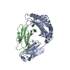
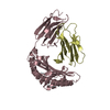
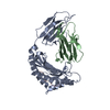
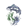
Database: PDB / ID: 4z76
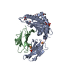
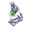
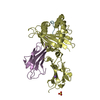
Title
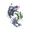
Weak TCR binding to an unstable insulin epitope drives type 1 diabetes
Components
Beta-2-microglobulin
H-2 class I histocompatibility antigen, K-D alpha chain
Insulin
Keywords
IMMUNE SYSTEM / Immunoglobulin / H-2Kd / Type 1 Diabetes
Function / homology
Function and homology information
negative regulation of glycogen catabolic process / : / negative regulation of fatty acid metabolic process / Signaling by Insulin receptor / IRS activation / Insulin processing / regulation of protein secretion / positive regulation of peptide hormone secretion / negative regulation of feeding behavior / negative regulation of acute inflammatory response ...negative regulation of glycogen catabolic process / : / negative regulation of fatty acid metabolic process / Signaling by Insulin receptor / IRS activation / Insulin processing / regulation of protein secretion / positive regulation of peptide hormone secretion / negative regulation of feeding behavior / negative regulation of acute inflammatory response / Regulation of gene expression in beta cells / positive regulation of respiratory burst / alpha-beta T cell activation / Synthesis, secretion, and deacylation of Ghrelin / negative regulation of protein secretion / positive regulation of dendritic spine maintenance / negative regulation of gluconeogenesis / fatty acid homeostasis / positive regulation of glycogen biosynthetic process / positive regulation of insulin receptor signaling pathway / Signal attenuation / FOXO-mediated transcription of oxidative stress, metabolic and neuronal genes / negative regulation of lipid catabolic process / negative regulation of respiratory burst involved in inflammatory response / antigen processing and presentation of peptide antigen via MHC class I / positive regulation of lipid biosynthetic process / negative regulation of oxidative stress-induced intrinsic apoptotic signaling pathway / nitric oxide-cGMP-mediated signaling / regulation of protein localization to plasma membrane / positive regulation of nitric-oxide synthase activity / transport vesicle / Insulin receptor recycling / COPI-mediated anterograde transport / negative regulation of reactive oxygen species biosynthetic process / positive regulation of brown fat cell differentiation / insulin-like growth factor receptor binding / NPAS4 regulates expression of target genes / neuron projection maintenance / positive regulation of mitotic nuclear division / endoplasmic reticulum-Golgi intermediate compartment membrane / Insulin receptor signalling cascade / early endosome lumen / Nef mediated downregulation of MHC class I complex cell surface expression / DAP12 interactions / positive regulation of glycolytic process / endosome lumen / acute-phase response / positive regulation of cytokine production / positive regulation of D-glucose import across plasma membrane / T cell mediated cytotoxicity / Endosomal/Vacuolar pathway / insulin receptor binding / positive regulation of long-term synaptic potentiation / positive regulation of protein secretion / Antigen Presentation: Folding, assembly and peptide loading of class I MHC / lumenal side of endoplasmic reticulum membrane / regulation of iron ion transport / positive regulation of cell differentiation / cellular response to iron(III) ion / antigen processing and presentation of exogenous protein antigen via MHC class Ib, TAP-dependent / wound healing / negative regulation of iron ion transport / negative regulation of forebrain neuron differentiation / regulation of erythrocyte differentiation / peptide antigen assembly with MHC class I protein complex / ER to Golgi transport vesicle membrane / response to molecule of bacterial origin / HFE-transferrin receptor complex / Regulation of insulin secretion / MHC class I peptide loading complex / transferrin transport / positive regulation of neuron projection development / negative regulation of receptor-mediated endocytosis / cellular response to iron ion / hormone activity / positive regulation of T cell cytokine production / antigen processing and presentation of endogenous peptide antigen via MHC class I / negative regulation of protein catabolic process / MHC class I protein complex / peptide antigen assembly with MHC class II protein complex / positive regulation of protein localization to nucleus / negative regulation of neurogenesis / multicellular organismal-level iron ion homeostasis / cellular response to nicotine / MHC class II protein complex / positive regulation of receptor-mediated endocytosis / regulation of synaptic plasticity / positive regulation of T cell mediated cytotoxicity / glucose metabolic process / Golgi lumen / negative regulation of epithelial cell proliferation / specific granule lumen / antigen processing and presentation of exogenous peptide antigen via MHC class II / positive regulation of immune response / peptide antigen binding / cognition / vasodilation / phagocytic vesicle membrane / recycling endosome membrane / positive regulation of T cell activation Similarity search - Function
Insulin / Insulin family / Insulin-like / Insulin/IGF/Relaxin family / Insulin / insulin-like growth factor / relaxin family. / Insulin, conserved site / Insulin family signature. / Insulin-like superfamily / MHC class I-like antigen recognition-like / Murine Class I Major Histocompatibility Complex, H2-DB; Chain A, domain 1 ...Insulin / Insulin family / Insulin-like / Insulin/IGF/Relaxin family / Insulin / insulin-like growth factor / relaxin family. / Insulin, conserved site / Insulin family signature. / Insulin-like superfamily / MHC class I-like antigen recognition-like / Murine Class I Major Histocompatibility Complex, H2-DB; Chain A, domain 1 / MHC class I alpha chain, alpha1 alpha2 domains / Class I Histocompatibility antigen, domains alpha 1 and 2 / Beta-2-Microglobulin / : / MHC class I-like antigen recognition-like / MHC class I-like antigen recognition-like superfamily / MHC classes I/II-like antigen recognition protein / : / Immunoglobulin/major histocompatibility complex, conserved site / Immunoglobulins and major histocompatibility complex proteins signature. / Immunoglobulin C-Type / Immunoglobulin C1-set / Immunoglobulin C1-set domain / Ig-like domain profile. / Immunoglobulin-like domain / Immunoglobulin-like domain superfamily / Immunoglobulin-like fold / Immunoglobulins / Immunoglobulin-like / Sandwich / 2-Layer Sandwich / Mainly Beta / Alpha Beta Similarity search - Domain/homology
Insulin / H-2 class I histocompatibility antigen, K-D alpha chain / Beta-2-microglobulin Similarity search - Component
Mass: 18.015 Da / Num. of mol.: 465 / Source method: isolated from a natural source / Formula: H2O
-
Details
Has protein modification
Y
-
Experimental details
-
Experiment
Experiment
Method: X-RAY DIFFRACTION
-
Sample preparation
Crystal
Density Matthews: 2.24 Å3/Da / Density % sol: 44.99 %
Crystal grow
Temperature: 291 K / Method: vapor diffusion / pH: 8 Details: G9V crystals were grown in 20% PEG 6000, 0.2 M calcium chloride, 0.1 M Tris propane pH 8.0
Method to determine structure: MOLECULAR REPLACEMENT / Resolution: 1.88→38.268 Å / Cor.coef. Fo:Fc: 0.957 / Cor.coef. Fo:Fc free: 0.933 / WRfactor Rfree: 0.2169 / WRfactor Rwork: 0.1721 / FOM work R set: 0.8159 / SU B: 9.281 / SU ML: 0.134 / SU R Cruickshank DPI: 0.1906 / SU Rfree: 0.1647 / Cross valid method: THROUGHOUT / σ(F): 0 / ESU R: 0.191 / ESU R Free: 0.165 / Stereochemistry target values: MAXIMUM LIKELIHOOD Details: HYDROGENS HAVE BEEN ADDED IN THE RIDING POSITIONS U VALUES : WITH TLS ADDED
Rfactor
Num. reflection
% reflection
Selection details
Rfree
0.2296
2886
5.1 %
RANDOM
Rwork
0.184
-
-
-
obs
0.1863
54089
89.27 %
-
Solvent computation
Ion probe radii: 0.8 Å / Shrinkage radii: 0.8 Å / VDW probe radii: 1.2 Å / Solvent model: MASK
In the structure databanks used in Yorodumi, some data are registered as the other names, "COVID-19 virus" and "2019-nCoV". Here are the details of the virus and the list of structure data.
Jan 31, 2019. EMDB accession codes are about to change! (news from PDBe EMDB page)
EMDB accession codes are about to change! (news from PDBe EMDB page)
The allocation of 4 digits for EMDB accession codes will soon come to an end. Whilst these codes will remain in use, new EMDB accession codes will include an additional digit and will expand incrementally as the available range of codes is exhausted. The current 4-digit format prefixed with “EMD-” (i.e. EMD-XXXX) will advance to a 5-digit format (i.e. EMD-XXXXX), and so on. It is currently estimated that the 4-digit codes will be depleted around Spring 2019, at which point the 5-digit format will come into force.
The EM Navigator/Yorodumi systems omit the EMD- prefix.
Related info.:Q: What is EMD? / ID/Accession-code notation in Yorodumi/EM Navigator
Yorodumi is a browser for structure data from EMDB, PDB, SASBDB, etc.
This page is also the successor to EM Navigator detail page, and also detail information page/front-end page for Omokage search.
The word "yorodu" (or yorozu) is an old Japanese word meaning "ten thousand". "mi" (miru) is to see.
Related info.:EMDB / PDB / SASBDB / Comparison of 3 databanks / Yorodumi Search / Aug 31, 2016. New EM Navigator & Yorodumi / Yorodumi Papers / Jmol/JSmol / Function and homology information / Changes in new EM Navigator and Yorodumi
 Movie
Movie Controller

 Yorodumi
 Open data
 Basic information
 Components
 Keywords
 Function and homology information
 Homo sapiens (human)
 X-RAY DIFFRACTION /
  Authors
 Citation
 Structure visualization
 Downloads & links
 Other downloads
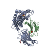


 PDBj
PDBj


 Assembly


 Mass: 62.068 Da / Num. of mol.: 6 / Source method: obtained synthetically / Formula: C2H6O2
 Mass: 92.094 Da / Num. of mol.: 3 / Source method: obtained synthetically / Formula: C3H8O3
 Mass: 96.063 Da / Num. of mol.: 15 / Source method: obtained synthetically / Formula: SO4
 Sample preparation
 / Beamline: I04-1 / Wavelength: 0.9173 Å
 Processing