 Movie
Movie Controller


+ Open data

- Basic information
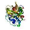
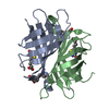
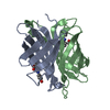
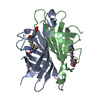
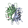
| Entry | Database: PDB / ID: 4z27 | ||||||
|---|---|---|---|---|---|---|---|
| Title | Crystal structure of apo short hoefavidin | ||||||
 Components Components | Avidin family | ||||||
 Keywords Keywords | Biotin-binding protein / high affinity system / bacterial avidins | ||||||
| Function / homology |  Function and homology information Function and homology informationbiotin binding / protein homodimerization activity / extracellular region Similarity search - Function | ||||||
| Biological species |  Hoeflea phototrophica DFL-43 (bacteria) Hoeflea phototrophica DFL-43 (bacteria) | ||||||
| Method |  X-RAY DIFFRACTION / SYNCHROTRON / MOLECULAR REPLACEMENT / Resolution: 1.34 Å X-RAY DIFFRACTION / SYNCHROTRON / MOLECULAR REPLACEMENT / Resolution: 1.34 Å | ||||||
 Authors Authors | Livnah, O. / Avraham, O. | ||||||
 Citation Citation | Journal: J.Struct.Biol. / Year: 2015 Title: Hoefavidin: A dimeric bacterial avidin with a C-terminal binding tail. Authors: Avraham, O. / Meir, A. / Fish, A. / Bayer, E.A. / Livnah, O. | ||||||
| History |
|
- Structure visualization
| Structure viewer | Molecule: MolmilJmol/JSmol |
|---|
- Downloads & links
-Download
| PDBx/mmCIF format | 4z27.cif.gz | 109.6 KB | Display | PDBx/mmCIF format |
|---|---|---|---|---|
| PDB format | pdb4z27.ent.gz | 85.1 KB | Display | PDB format |
| PDBx/mmJSON format | 4z27.json.gz | Tree view | PDBx/mmJSON format | |
| Others |  Other downloads Other downloads |
-Validation report
| Arichive directory | https://data.pdbj.org/pub/pdb/validation_reports/z2/4z27ftp://data.pdbj.org/pub/pdb/validation_reports/z2/4z27 | HTTPS FTP |
|---|
-Related structure data
| Related structure data |  4z28C 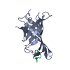 4z2oC 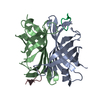 4z2pC 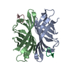 4z2vC 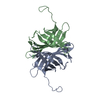 4z6jSC S: Starting model for refinement C: citing same article ( |
|---|---|
| Similar structure data |


-Links
 PDBj
PDBj- Assembly
| Deposited unit | 
| ||||||||
|---|---|---|---|---|---|---|---|---|---|
| 1 |
| ||||||||
| Unit cell |
|
-Components
| #1: Protein | Mass: 14341.508 Da / Num. of mol.: 2 / Fragment: UNP residues 21-154 Source method: isolated from a genetically manipulated source Source: (gene. exp.) Hoeflea phototrophica DFL-43 (bacteria)Gene: HPDFL43_17171 / Production host: #2: Water | ChemComp-HOH / |  Mass: 18.015 Da / Num. of mol.: 115 / Source method: isolated from a natural source / Formula: H2O Mass: 18.015 Da / Num. of mol.: 115 / Source method: isolated from a natural source / Formula: H2OHas protein modification | Y | |
|---|
-Experimental details
-Experiment
| Experiment | Method: X-RAY DIFFRACTION |
|---|
- Sample preparation
| Crystal | Density Matthews: 2.72 Å3/Da / Density % sol: 54.73 % |
|---|---|
| Crystal grow | Temperature: 293 K / Method: vapor diffusion, sitting drop Details: Streak seeding was applied with source crystals. Crystals appeared within 24 hours and grew to their maximal size within 2 weeks |
-Data collection
| Diffraction | Mean temperature: 100 K |
|---|---|
| Diffraction source | Source: SYNCHROTRON / Site: ESRF  / Beamline: ID23-1 / Wavelength: 0.98 Å / Beamline: ID23-1 / Wavelength: 0.98 Å |
| Detector | Type: DECTRIS PILATUS 6M / Detector: PIXEL / Date: Sep 26, 2013 |
| Radiation | Protocol: SINGLE WAVELENGTH / Monochromatic (M) / Laue (L): M / Scattering type: x-ray |
| Radiation wavelength | Wavelength: 0.98 Å / Relative weight: 1 |
| Reflection | Resolution: 1.34→57.4 Å / Num. obs: 72099 / % possible obs: 95.6 % / Redundancy: 13.8 % / Rmerge(I) obs: 0.047 / Net I/σ(I): 27.1 |
| Reflection shell | Resolution: 1.34→1.39 Å / Rmerge(I) obs: 0.564 |
- Processing
| Software |
| ||||||||||||||||||||||||||||||||||||||||||||||||||||||||||||||||||||||||||||||||||||||||||||||||||||||||||||||||||||||||||||||||||||||||||||||||||||||||||||||||||||||||||||||||||||||
|---|---|---|---|---|---|---|---|---|---|---|---|---|---|---|---|---|---|---|---|---|---|---|---|---|---|---|---|---|---|---|---|---|---|---|---|---|---|---|---|---|---|---|---|---|---|---|---|---|---|---|---|---|---|---|---|---|---|---|---|---|---|---|---|---|---|---|---|---|---|---|---|---|---|---|---|---|---|---|---|---|---|---|---|---|---|---|---|---|---|---|---|---|---|---|---|---|---|---|---|---|---|---|---|---|---|---|---|---|---|---|---|---|---|---|---|---|---|---|---|---|---|---|---|---|---|---|---|---|---|---|---|---|---|---|---|---|---|---|---|---|---|---|---|---|---|---|---|---|---|---|---|---|---|---|---|---|---|---|---|---|---|---|---|---|---|---|---|---|---|---|---|---|---|---|---|---|---|---|---|---|---|---|---|
| Refinement | Method to determine structure: MOLECULAR REPLACEMENT Starting model: 4Z6J Resolution: 1.34→57.42 Å / Cor.coef. Fo:Fc: 0.971 / Cor.coef. Fo:Fc free: 0.97 / SU B: 1.793 / SU ML: 0.032 / Cross valid method: THROUGHOUT / ESU R: 0.047 / ESU R Free: 0.046 / Stereochemistry target values: MAXIMUM LIKELIHOOD / Details: HYDROGENS HAVE BEEN ADDED IN THE RIDING POSITIONS
| ||||||||||||||||||||||||||||||||||||||||||||||||||||||||||||||||||||||||||||||||||||||||||||||||||||||||||||||||||||||||||||||||||||||||||||||||||||||||||||||||||||||||||||||||||||||
| Solvent computation | Ion probe radii: 0.8 Å / Shrinkage radii: 0.8 Å / VDW probe radii: 1.2 Å / Solvent model: MASK | ||||||||||||||||||||||||||||||||||||||||||||||||||||||||||||||||||||||||||||||||||||||||||||||||||||||||||||||||||||||||||||||||||||||||||||||||||||||||||||||||||||||||||||||||||||||
| Displacement parameters | Biso mean: 22.422 Å2
| ||||||||||||||||||||||||||||||||||||||||||||||||||||||||||||||||||||||||||||||||||||||||||||||||||||||||||||||||||||||||||||||||||||||||||||||||||||||||||||||||||||||||||||||||||||||
| Refinement step | Cycle: LAST / Resolution: 1.34→57.42 Å
| ||||||||||||||||||||||||||||||||||||||||||||||||||||||||||||||||||||||||||||||||||||||||||||||||||||||||||||||||||||||||||||||||||||||||||||||||||||||||||||||||||||||||||||||||||||||
| Refine LS restraints |
|