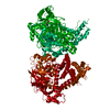
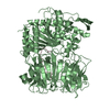
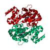
Entry Database : PDB / ID : 4y06Title Crystal structure of the DAP BII (G675R) dipeptide complex Dipeptidyl aminopeptidase BII Keywords Function / homology Function Domain/homology Component
/ / / / / Biological species Pseudoxanthomonas mexicana (bacteria)Method / / / Resolution : 2.18 Å Authors Sakamoto, Y. / Iizuka, I. / Tateoka, C. / Roppongi, S. / Fujimoto, M. / Nonaka, T. / Ogasawara, W. / Tanaka, N. Funding support Organization Grant number Country Japan Society for the Promotion of Science 25462872
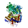
Journal : Sci Rep / Year : 2015Title : Structural and mutational analyses of dipeptidyl peptidase 11 from Porphyromonas gingivalis reveal the molecular basis for strict substrate specificity.Authors : Sakamoto, Y. / Suzuki, Y. / Iizuka, I. / Tateoka, C. / Roppongi, S. / Fujimoto, M. / Inaka, K. / Tanaka, H. / Yamada, M. / Ohta, K. / Gouda, H. / Nonaka, T. / Ogasawara, W. / Tanaka, N. History Deposition Feb 5, 2015 Deposition site / Processing site Revision 1.0 Jul 15, 2015 Provider / Type Revision 1.1 Feb 5, 2020 Group / Derived calculations / Source and taxonomyCategory / entity_src_gen / pdbx_struct_oper_listItem / _entity_src_gen.pdbx_alt_source_flag / _pdbx_struct_oper_list.symmetry_operationRevision 1.2 Nov 8, 2023 Group Data collection / Database references ... Data collection / Database references / Derived calculations / Refinement description Category chem_comp_atom / chem_comp_bond ... chem_comp_atom / chem_comp_bond / database_2 / pdbx_initial_refinement_model / pdbx_struct_conn_angle / struct_conn / struct_conn_type Item _database_2.pdbx_DOI / _database_2.pdbx_database_accession ... _database_2.pdbx_DOI / _database_2.pdbx_database_accession / _pdbx_struct_conn_angle.ptnr1_auth_asym_id / _pdbx_struct_conn_angle.ptnr1_auth_comp_id / _pdbx_struct_conn_angle.ptnr1_auth_seq_id / _pdbx_struct_conn_angle.ptnr1_label_asym_id / _pdbx_struct_conn_angle.ptnr1_label_atom_id / _pdbx_struct_conn_angle.ptnr1_label_comp_id / _pdbx_struct_conn_angle.ptnr1_label_seq_id / _pdbx_struct_conn_angle.ptnr1_symmetry / _pdbx_struct_conn_angle.ptnr2_auth_asym_id / _pdbx_struct_conn_angle.ptnr2_auth_seq_id / _pdbx_struct_conn_angle.ptnr2_label_asym_id / _pdbx_struct_conn_angle.ptnr2_symmetry / _pdbx_struct_conn_angle.ptnr3_auth_asym_id / _pdbx_struct_conn_angle.ptnr3_auth_comp_id / _pdbx_struct_conn_angle.ptnr3_auth_seq_id / _pdbx_struct_conn_angle.ptnr3_label_asym_id / _pdbx_struct_conn_angle.ptnr3_label_atom_id / _pdbx_struct_conn_angle.ptnr3_label_comp_id / _pdbx_struct_conn_angle.ptnr3_label_seq_id / _pdbx_struct_conn_angle.ptnr3_symmetry / _pdbx_struct_conn_angle.value / _struct_conn.conn_type_id / _struct_conn.id / _struct_conn.pdbx_dist_value / _struct_conn.pdbx_leaving_atom_flag / _struct_conn.pdbx_value_order / _struct_conn.ptnr1_auth_asym_id / _struct_conn.ptnr1_auth_comp_id / _struct_conn.ptnr1_auth_seq_id / _struct_conn.ptnr1_label_asym_id / _struct_conn.ptnr1_label_atom_id / _struct_conn.ptnr1_label_comp_id / _struct_conn.ptnr1_label_seq_id / _struct_conn.ptnr1_symmetry / _struct_conn.ptnr2_auth_asym_id / _struct_conn.ptnr2_auth_comp_id / _struct_conn.ptnr2_auth_seq_id / _struct_conn.ptnr2_label_asym_id / _struct_conn.ptnr2_label_atom_id / _struct_conn.ptnr2_label_comp_id / _struct_conn.ptnr2_label_seq_id / _struct_conn.ptnr2_symmetry / _struct_conn_type.id Revision 1.3 Nov 6, 2024 Group / Category / pdbx_modification_feature
Show all Show less
 Movie
Movie Controller


 Open data
 Basic information
 Components
 Keywords
 Function and homology information
 Pseudoxanthomonas mexicana (bacteria)
 X-RAY DIFFRACTION /
  Authors
 Japan, 1items
  Citation
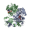
 Structure visualization
 Downloads & links
 Other downloads
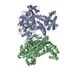


 PDBj
PDBj


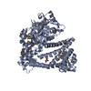
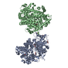
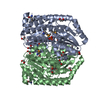
 Assembly


 Type: L-peptide linking / Mass: 131.173 Da / Num. of mol.: 2 / Source method: obtained synthetically / Formula: C6H13NO2
 Type: L-peptide linking / Mass: 147.129 Da / Num. of mol.: 2 / Source method: obtained synthetically / Formula: C5H9NO4
 Mass: 92.094 Da / Num. of mol.: 14 / Source method: obtained synthetically / Formula: C3H8O3
 Mass: 65.409 Da / Num. of mol.: 5 / Source method: obtained synthetically / Formula: Zn
 Sample preparation
 Processing