 Movie
Movie Controller

[English] 日本語
 Yorodumi
- PDB-2bkl: Structural and Mechanistic Analysis of Two Prolyl Endopeptidases:... -
+ Open data

- Basic information
| Entry | Database: PDB / ID: 2bkl | ||||||
|---|---|---|---|---|---|---|---|
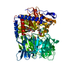
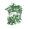
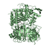
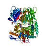
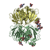
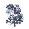
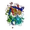
| Title | Structural and Mechanistic Analysis of Two Prolyl Endopeptidases: Role of Inter-Domain Dynamics in Catalysis and Specificity | ||||||
 Components Components | PROLYL ENDOPEPTIDASE | ||||||
 Keywords Keywords | HYDROLASE / PROLYL ENDOPEPTIDASE / MECHANISTIC STUDY / CELIAC SPRUE / PROTEASE | ||||||
| Function / homology |  Function and homology information Function and homology informationprolyl oligopeptidase / oligopeptidase activity / serine-type endopeptidase activity / proteolysis / cytosol Similarity search - Function | ||||||
| Biological species |  MYXOCOCCUS XANTHUS (bacteria) MYXOCOCCUS XANTHUS (bacteria) | ||||||
| Method |  X-RAY DIFFRACTION / SYNCHROTRON / MOLECULAR REPLACEMENT / Resolution: 1.5 Å X-RAY DIFFRACTION / SYNCHROTRON / MOLECULAR REPLACEMENT / Resolution: 1.5 Å | ||||||
 Authors Authors | Khosla, C. / Shan, L. / Mathews, I.I. | ||||||
 Citation Citation | Journal: Proc.Natl.Acad.Sci.USA / Year: 2005 Title: Structural and Mechanistic Analysis of Two Prolyl Endopeptidases: Role of Interdomain Dynamics in Catalysis and Specificity Authors: Shan, L. / Mathews, I.I. / Khosla, C. #1: Journal: Biochem.J. / Year: 2004 Title: Comparative Biochemical Analysis of Three Bacterial Prolyl Endopeptidases: Implications for Celiac Sprue Authors: Shan, L. / Marti, T. / Sollid, L.M. / Gray, G.M. / Khosla, C. #2: Journal: Science / Year: 2002 Title: Structural Basis for Gluten Intolerance in Celiac Sprue Authors: Shan, L. / Molberg, O. / Parrot, I. / Hausch, F. / Filiz, F. / Gray, G.M. / Sollid, L.M. / Khosla, C. #3: Journal: Cell.Mol.Life.Sci. / Year: 2002 Title: The Prolyl Oligopeptidase Family Authors: Polgar, L. | ||||||
| History |
| ||||||
| Remark 700 | SHEET THE SHEET STRUCTURE OF THIS MOLECULE IS BIFURCATED. IN ORDER TO REPRESENT THIS FEATURE IN ... SHEET THE SHEET STRUCTURE OF THIS MOLECULE IS BIFURCATED. IN ORDER TO REPRESENT THIS FEATURE IN THE SHEET RECORDS BELOW, TWO SHEETS ARE DEFINED. |
- Structure visualization
| Structure viewer | Molecule: MolmilJmol/JSmol |
|---|
- Downloads & links
-Download
| PDBx/mmCIF format | 2bkl.cif.gz | 303.3 KB | Display | PDBx/mmCIF format |
|---|---|---|---|---|
| PDB format | pdb2bkl.ent.gz | 244.9 KB | Display | PDB format |
| PDBx/mmJSON format | 2bkl.json.gz | Tree view | PDBx/mmJSON format | |
| Others |  Other downloads Other downloads |
-Validation report
| Arichive directory | https://data.pdbj.org/pub/pdb/validation_reports/bk/2bklftp://data.pdbj.org/pub/pdb/validation_reports/bk/2bkl | HTTPS FTP |
|---|
-Related structure data
| Related structure data |  1yr2C  1qfsS S: Starting model for refinement C: citing same article ( |
|---|---|
| Similar structure data |


-Links
 PDBj
PDBj
- Assembly
| Deposited unit | 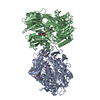
| ||||||||
|---|---|---|---|---|---|---|---|---|---|
| 1 | 
| ||||||||
| 2 | 
| ||||||||
| Unit cell |
|
-Components
| #1: Protein | Mass: 77777.359 Da / Num. of mol.: 2 Source method: isolated from a genetically manipulated source Details: Z-ALA PROLINAL / Source: (gene. exp.) MYXOCOCCUS XANTHUS (bacteria) / Plasmid: PET28B / Production host: #2: Chemical |   Mass: 320.340 Da / Num. of mol.: 3 / Source method: obtained synthetically / Formula: C16H20N2O5 Mass: 320.340 Da / Num. of mol.: 3 / Source method: obtained synthetically / Formula: C16H20N2O5#3: Chemical |   Mass: 195.237 Da / Num. of mol.: 3 / Source method: obtained synthetically / Formula: C6H13NO4S / Comment: pH buffer*YM Mass: 195.237 Da / Num. of mol.: 3 / Source method: obtained synthetically / Formula: C6H13NO4S / Comment: pH buffer*YM#4: Chemical | ChemComp-SO4 / |   Mass: 96.063 Da / Num. of mol.: 1 / Source method: obtained synthetically / Formula: SO4 Mass: 96.063 Da / Num. of mol.: 1 / Source method: obtained synthetically / Formula: SO4#5: Water | ChemComp-HOH / |  Mass: 18.015 Da / Num. of mol.: 1371 / Source method: isolated from a natural source / Formula: H2O Mass: 18.015 Da / Num. of mol.: 1371 / Source method: isolated from a natural source / Formula: H2OHas protein modification | N | |
|---|
-Experimental details
-Experiment
| Experiment | Method: X-RAY DIFFRACTION / Number of used crystals: 1 |
|---|
- Sample preparation
| Crystal | Density Matthews: 2.3 Å3/Da / Density % sol: 45 % |
|---|---|
| Crystal grow | pH: 6 / Details: 26% METHOXY PEG 5K AND 0.1MES (PH 6.01). |
-Data collection
| Diffraction | Mean temperature: 100 K |
|---|---|
| Diffraction source | Source: SYNCHROTRON / Site: ALS  / Beamline: 8.2.2 / Wavelength: 1 / Beamline: 8.2.2 / Wavelength: 1 |
| Detector | Type: ADSC CCD / Detector: CCD / Date: Dec 19, 2003 |
| Radiation | Monochromator: DOUBLE CRYSTAL, SI(111) / Protocol: SINGLE WAVELENGTH / Monochromatic (M) / Laue (L): M / Scattering type: x-ray |
| Radiation wavelength | Wavelength: 1 Å / Relative weight: 1 |
| Reflection | Resolution: 1.5→50 Å / Num. obs: 225366 / % possible obs: 98.9 % / Observed criterion σ(I): 0 / Redundancy: 4.1 % / Biso Wilson estimate: 15.3 Å2 / Rmerge(I) obs: 0.07 / Net I/σ(I): 12.8 |
| Reflection shell | Resolution: 1.5→1.55 Å / Redundancy: 4 % / Rmerge(I) obs: 0.47 / Mean I/σ(I) obs: 2.8 / % possible all: 97.7 |
- Processing
| Software |
| ||||||||||||||||||||||||||||||||||||||||||||||||||||||||||||||||||||||||||||||||||||||||||||||||||||||||||||||||||||||||||||||||||||||||||||||||||||||||||||||||||||||||||||||||||||||
|---|---|---|---|---|---|---|---|---|---|---|---|---|---|---|---|---|---|---|---|---|---|---|---|---|---|---|---|---|---|---|---|---|---|---|---|---|---|---|---|---|---|---|---|---|---|---|---|---|---|---|---|---|---|---|---|---|---|---|---|---|---|---|---|---|---|---|---|---|---|---|---|---|---|---|---|---|---|---|---|---|---|---|---|---|---|---|---|---|---|---|---|---|---|---|---|---|---|---|---|---|---|---|---|---|---|---|---|---|---|---|---|---|---|---|---|---|---|---|---|---|---|---|---|---|---|---|---|---|---|---|---|---|---|---|---|---|---|---|---|---|---|---|---|---|---|---|---|---|---|---|---|---|---|---|---|---|---|---|---|---|---|---|---|---|---|---|---|---|---|---|---|---|---|---|---|---|---|---|---|---|---|---|---|
| Refinement | Method to determine structure: MOLECULAR REPLACEMENT Starting model: PDB ENTRY 1QFS Resolution: 1.5→30 Å / Cor.coef. Fo:Fc: 0.966 / Cor.coef. Fo:Fc free: 0.958 / SU B: 1.062 / SU ML: 0.04 / Cross valid method: THROUGHOUT / σ(F): 0 / ESU R: 0.066 / ESU R Free: 0.067 / Stereochemistry target values: MAXIMUM LIKELIHOOD Details: HYDROGENS HAVE BEEN ADDED IN THE RIDING POSITIONS. SOME OF THE RESIDUES ARE MODELED WITH ALTERNATE CONFORMATIONS.
| ||||||||||||||||||||||||||||||||||||||||||||||||||||||||||||||||||||||||||||||||||||||||||||||||||||||||||||||||||||||||||||||||||||||||||||||||||||||||||||||||||||||||||||||||||||||
| Solvent computation | Ion probe radii: 0.8 Å / Shrinkage radii: 0.8 Å / VDW probe radii: 1.4 Å / Solvent model: BABINET MODEL WITH MASK | ||||||||||||||||||||||||||||||||||||||||||||||||||||||||||||||||||||||||||||||||||||||||||||||||||||||||||||||||||||||||||||||||||||||||||||||||||||||||||||||||||||||||||||||||||||||
| Displacement parameters | Biso mean: 10.34 Å2
| ||||||||||||||||||||||||||||||||||||||||||||||||||||||||||||||||||||||||||||||||||||||||||||||||||||||||||||||||||||||||||||||||||||||||||||||||||||||||||||||||||||||||||||||||||||||
| Refinement step | Cycle: LAST / Resolution: 1.5→30 Å
| ||||||||||||||||||||||||||||||||||||||||||||||||||||||||||||||||||||||||||||||||||||||||||||||||||||||||||||||||||||||||||||||||||||||||||||||||||||||||||||||||||||||||||||||||||||||
| Refine LS restraints |
|