- PDB-4xxb: Crystal structure of human MDM2-RPL11 -
+
Open data
ID or keywords:
Loading...
-
Basic information
Entry
Database: PDB / ID: 4xxb
Title
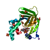
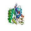
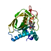
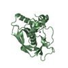
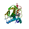
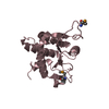
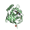
Crystal structure of human MDM2-RPL11
Components
60S ribosomal protein L11
E3 ubiquitin-protein ligase Mdm2
Keywords
RNA BINDING PROTEIN/METAL BINDING PROTEIN / MDM2 / RPL11 / ModPipe Model of UP / RNA BINDING PROTEIN-METAL BINDING PROTEIN complex
Function / homology
Function and homology information
cellular response to vitamin B1 / response to formaldehyde / response to water-immersion restraint stress / response to ether / traversing start control point of mitotic cell cycle / atrial septum development / regulation of protein catabolic process at postsynapse, modulating synaptic transmission / fibroblast activation / Trafficking of AMPA receptors / receptor serine/threonine kinase binding ...cellular response to vitamin B1 / response to formaldehyde / response to water-immersion restraint stress / response to ether / traversing start control point of mitotic cell cycle / atrial septum development / regulation of protein catabolic process at postsynapse, modulating synaptic transmission / fibroblast activation / Trafficking of AMPA receptors / receptor serine/threonine kinase binding / negative regulation of protein neddylation / peroxisome proliferator activated receptor binding / negative regulation of intrinsic apoptotic signaling pathway by p53 class mediator / positive regulation of vascular associated smooth muscle cell migration / negative regulation of protein processing / SUMO transferase activity / response to steroid hormone / NEDD8 ligase activity / AKT phosphorylates targets in the cytosol / response to iron ion / atrioventricular valve morphogenesis / endocardial cushion morphogenesis / cellular response to peptide hormone stimulus / ventricular septum development / negative regulation of ubiquitin protein ligase activity / positive regulation of muscle cell differentiation / cardiac septum morphogenesis / regulation of postsynaptic neurotransmitter receptor internalization / blood vessel development / SUMOylation of ubiquitinylation proteins / ligase activity / cellular response to alkaloid / Constitutive Signaling by AKT1 E17K in Cancer / regulation of protein catabolic process / negative regulation of signal transduction by p53 class mediator / negative regulation of DNA damage response, signal transduction by p53 class mediator / SUMOylation of transcription factors / Peptide chain elongation / Selenocysteine synthesis / Formation of a pool of free 40S subunits / response to magnesium ion / cellular response to UV-C / Eukaryotic Translation Termination / protein sumoylation / ubiquitin ligase inhibitor activity / cellular response to actinomycin D / Response of EIF2AK4 (GCN2) to amino acid deficiency / SRP-dependent cotranslational protein targeting to membrane / negative regulation of ubiquitin-dependent protein catabolic process / positive regulation of signal transduction by p53 class mediator / Viral mRNA Translation / blood vessel remodeling / cellular response to estrogen stimulus / Nonsense Mediated Decay (NMD) independent of the Exon Junction Complex (EJC) / GTP hydrolysis and joining of the 60S ribosomal subunit / protein localization to nucleus / L13a-mediated translational silencing of Ceruloplasmin expression / positive regulation of protein binding / Major pathway of rRNA processing in the nucleolus and cytosol / ribonucleoprotein complex binding / protein targeting / Nonsense Mediated Decay (NMD) enhanced by the Exon Junction Complex (EJC) / protein autoubiquitination / positive regulation of vascular associated smooth muscle cell proliferation / NPAS4 regulates expression of target genes / negative regulation of proteasomal ubiquitin-dependent protein catabolic process / transcription repressor complex / cytosolic ribosome / positive regulation of mitotic cell cycle / regulation of heart rate / proteolysis involved in protein catabolic process / regulation of signal transduction by p53 class mediator / positive regulation of protein export from nucleus / ribosomal large subunit biogenesis / ubiquitin binding / response to cocaine / DNA damage response, signal transduction by p53 class mediator / Stabilization of p53 / establishment of protein localization / Regulation of RUNX3 expression and activity / cellular response to gamma radiation / Oncogene Induced Senescence / RING-type E3 ubiquitin transferase / protein destabilization / Regulation of TP53 Activity through Methylation / cellular response to growth factor stimulus / response to toxic substance / centriolar satellite / Regulation of expression of SLITs and ROBOs / cellular response to hydrogen peroxide / protein polyubiquitination / rRNA processing / ubiquitin-protein transferase activity / disordered domain specific binding / endocytic vesicle membrane / p53 binding / ubiquitin protein ligase activity / Signaling by ALK fusions and activated point mutants / Regulation of TP53 Degradation / positive regulation of proteasomal ubiquitin-dependent protein catabolic process Similarity search - Function
Zn-finger domain of Sec23/24 / E3 ubiquitin-protein ligase Mdm2 / MDM2, modified RING finger, HC subclass / p53 negative regulator Mdm2/Mdm4 / SWIB/MDM2 domain / SWIB/MDM2 domain / SWIB/MDM2 domain profile. / SWIB/MDM2 domain superfamily / Zn-finger in Ran binding protein and others / Zinc finger, C3HC4 type (RING finger) ...Zn-finger domain of Sec23/24 / E3 ubiquitin-protein ligase Mdm2 / MDM2, modified RING finger, HC subclass / p53 negative regulator Mdm2/Mdm4 / SWIB/MDM2 domain / SWIB/MDM2 domain / SWIB/MDM2 domain profile. / SWIB/MDM2 domain superfamily / Zn-finger in Ran binding protein and others / Zinc finger, C3HC4 type (RING finger) / Zinc finger RanBP2 type profile. / Zinc finger, RanBP2-type superfamily / Zinc finger RanBP2-type signature. / Zinc finger, RanBP2-type / Zinc finger RING-type profile. / SH3 type barrels. / Zinc finger, RING-type / Ribosomal protein L5, conserved site / Ribosomal protein L5 signature. / Ribosomal protein L5, N-terminal / Ribosomal protein L5 / Ribosomal protein L5, C-terminal / ribosomal L5P family C-terminus / Ribosomal protein L5 / Ribosomal protein L5 domain superfamily / Zinc finger, RING/FYVE/PHD-type / Roll / Mainly Beta Similarity search - Domain/homology
BETA-MERCAPTOETHANOL / IMIDAZOLE / Large ribosomal subunit protein uL5 / E3 ubiquitin-protein ligase Mdm2 Similarity search - Component
Resolution: 2.4→50 Å / Cor.coef. Fo:Fc: 0.959 / Cor.coef. Fo:Fc free: 0.941 / SU B: 8.084 / SU ML: 0.183 / Cross valid method: THROUGHOUT / ESU R: 0.396 / ESU R Free: 0.238 / Stereochemistry target values: MAXIMUM LIKELIHOOD / Details: HYDROGENS HAVE BEEN USED IF PRESENT IN THE INPUT
Rfactor
Num. reflection
% reflection
Selection details
Rfree
0.22654
447
4.8 %
RANDOM
Rwork
0.19202
-
-
-
obs
0.19366
8807
98.95 %
-
Solvent computation
Ion probe radii: 0.8 Å / Shrinkage radii: 0.8 Å / VDW probe radii: 1.2 Å / Solvent model: MASK
 Movie
Movie Controller


 Open data
 Basic information
 Components
 Keywords
 Function and homology information
 Homo sapiens (human)
 X-RAY DIFFRACTION /
  Authors
 Citation
 Structure visualization
 Downloads & links
 Other downloads
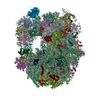


 PDBj
PDBj


 Assembly
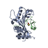


 Mass: 69.085 Da / Num. of mol.: 1 / Source method: obtained synthetically / Formula: C3H5N2
 Mass: 78.133 Da / Num. of mol.: 1 / Source method: obtained synthetically / Formula: C2H6OS
 Mass: 65.409 Da / Num. of mol.: 2 / Source method: obtained synthetically / Formula: Zn
 Sample preparation
 / Beamline: AR-NE3A / Wavelength: 1 Å
 Processing