 Movie
Movie Controller

[English] 日本語
 Yorodumi
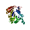
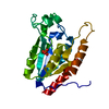
- PDB-3ikb: The structure of a conserved protein from Streptococcus mutans UA159. -
+ Open data

- Basic information
| Entry | Database: PDB / ID: 3ikb | ||||||
|---|---|---|---|---|---|---|---|
| Title | The structure of a conserved protein from Streptococcus mutans UA159. | ||||||
 Components Components | uncharacterized conserved protein | ||||||
 Keywords Keywords | structural genomics / unknown function / APC63946 / conserved protein / Streptococcus mutans UA159 / PSI-2 / protein structure initiative / midwest center for structural genomics / MCSG | ||||||
| Function / homology |  Function and homology information Function and homology information: / Uracil DNA glycosylase superfamily / UreE urease accessory protein, C-terminal domain / Uracil-DNA Glycosylase, subunit E / Uracil-DNA glycosylase-like domain / Uracil-DNA glycosylase-like / Uracil DNA glycosylase superfamily / Uracil-DNA glycosylase-like domain superfamily / 3-Layer(aba) Sandwich / Alpha Beta Similarity search - Domain/homology | ||||||
| Biological species |  Streptococcus mutans (bacteria) Streptococcus mutans (bacteria) | ||||||
| Method |  X-RAY DIFFRACTION / SYNCHROTRON / SAD / Resolution: 1.67 Å X-RAY DIFFRACTION / SYNCHROTRON / SAD / Resolution: 1.67 Å | ||||||
 Authors Authors | Tan, K. / Hatzos, C. / Buck, K. / Joachimiak, A. / Midwest Center for Structural Genomics (MCSG) | ||||||
 Citation Citation | Journal: To be Published Title: The structure of a conserved protein from Streptococcus mutans UA159. Authors: Tan, K. / Hatzos, C. / Buck, K. / Joachimiak, A. | ||||||
| History |
|
- Structure visualization
| Structure viewer | Molecule: MolmilJmol/JSmol |
|---|
- Downloads & links
-Download
| PDBx/mmCIF format | 3ikb.cif.gz | 105.3 KB | Display | PDBx/mmCIF format |
|---|---|---|---|---|
| PDB format | pdb3ikb.ent.gz | 81.6 KB | Display | PDB format |
| PDBx/mmJSON format | 3ikb.json.gz | Tree view | PDBx/mmJSON format | |
| Others |  Other downloads Other downloads |
-Validation report
| Arichive directory | https://data.pdbj.org/pub/pdb/validation_reports/ik/3ikbftp://data.pdbj.org/pub/pdb/validation_reports/ik/3ikb | HTTPS FTP |
|---|
-Related structure data
| Similar structure data | |
|---|---|
| Other databases |


-Links
 PDBj
PDBj


- Assembly
| Deposited unit | 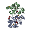
| ||||||||
|---|---|---|---|---|---|---|---|---|---|
| 1 | 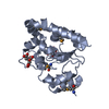
| ||||||||
| 2 | 
| ||||||||
| Unit cell |
| ||||||||
| Components on special symmetry positions |
| ||||||||
| Details | authors state that the biological unit is experimentally unknown. The protein is likely monomeric. |
-Components
| #1: Protein | Mass: 23141.729 Da / Num. of mol.: 2 Source method: isolated from a genetically manipulated source Source: (gene. exp.) Streptococcus mutans (bacteria) / Strain: UA159 / Gene: SMU_1238c, Streptococcus mutans / Plasmid: pMCSG19 / Production host: #2: Chemical |   Mass: 189.100 Da / Num. of mol.: 3 / Source method: obtained synthetically / Formula: C6H5O7 Mass: 189.100 Da / Num. of mol.: 3 / Source method: obtained synthetically / Formula: C6H5O7#3: Water | ChemComp-HOH / |  Mass: 18.015 Da / Num. of mol.: 450 / Source method: isolated from a natural source / Formula: H2O Mass: 18.015 Da / Num. of mol.: 450 / Source method: isolated from a natural source / Formula: H2OHas protein modification | Y | |
|---|
-Experimental details
-Experiment
| Experiment | Method: X-RAY DIFFRACTION / Number of used crystals: 1 |
|---|
- Sample preparation
| Crystal | Density Matthews: 2.35 Å3/Da / Density % sol: 47.74 % |
|---|---|
| Crystal grow | Temperature: 297 K / Method: vapor diffusion, sitting drop / pH: 5 Details: 0.1M Tris-sodium citrate, 20%w/v PEG6000, pH 5, VAPOR DIFFUSION, SITTING DROP, temperature 297K |
-Data collection
| Diffraction | Mean temperature: 100 K |
|---|---|
| Diffraction source | Source: SYNCHROTRON / Site: APS  / Beamline: 19-ID / Wavelength: 0.97937 Å / Beamline: 19-ID / Wavelength: 0.97937 Å |
| Detector | Type: ADSC QUANTUM 315 / Detector: CCD / Date: Mar 8, 2009 / Details: Mirror |
| Radiation | Monochromator: Si 111 crystal / Protocol: SINGLE WAVELENGTH / Monochromatic (M) / Laue (L): M / Scattering type: x-ray |
| Radiation wavelength | Wavelength: 0.97937 Å / Relative weight: 1 |
| Reflection | Resolution: 1.67→47 Å / Num. all: 51476 / Num. obs: 51476 / % possible obs: 97.6 % / Observed criterion σ(F): 0 / Observed criterion σ(I): 0 / Redundancy: 7.2 % / Rmerge(I) obs: 0.107 / Net I/σ(I): 32.7 |
| Reflection shell | Resolution: 1.67→1.7 Å / Redundancy: 7.3 % / Rmerge(I) obs: 0.873 / Mean I/σ(I) obs: 2.5 / Num. unique all: 2507 / % possible all: 97.5 |
- Processing
| Software |
| ||||||||||||||||||||||||||||||||||||||||||||||||||||||||||||||||||||||||||||||||||||||||||||||||||||||||||||||||||||||||||||||||||||||||||||||||||||||||||||||||||||||||||
|---|---|---|---|---|---|---|---|---|---|---|---|---|---|---|---|---|---|---|---|---|---|---|---|---|---|---|---|---|---|---|---|---|---|---|---|---|---|---|---|---|---|---|---|---|---|---|---|---|---|---|---|---|---|---|---|---|---|---|---|---|---|---|---|---|---|---|---|---|---|---|---|---|---|---|---|---|---|---|---|---|---|---|---|---|---|---|---|---|---|---|---|---|---|---|---|---|---|---|---|---|---|---|---|---|---|---|---|---|---|---|---|---|---|---|---|---|---|---|---|---|---|---|---|---|---|---|---|---|---|---|---|---|---|---|---|---|---|---|---|---|---|---|---|---|---|---|---|---|---|---|---|---|---|---|---|---|---|---|---|---|---|---|---|---|---|---|---|---|---|---|---|
| Refinement | Method to determine structure: SAD / Resolution: 1.67→46.93 Å / Cor.coef. Fo:Fc: 0.96 / Cor.coef. Fo:Fc free: 0.95 / SU B: 4.437 / SU ML: 0.068 / Cross valid method: THROUGHOUT / ESU R: 0.107 / ESU R Free: 0.103 / Stereochemistry target values: MAXIMUM LIKELIHOOD / Details: HYDROGENS HAVE BEEN ADDED IN THE RIDING POSITIONS
| ||||||||||||||||||||||||||||||||||||||||||||||||||||||||||||||||||||||||||||||||||||||||||||||||||||||||||||||||||||||||||||||||||||||||||||||||||||||||||||||||||||||||||
| Solvent computation | Ion probe radii: 0.8 Å / Shrinkage radii: 0.8 Å / VDW probe radii: 1.2 Å / Solvent model: MASK | ||||||||||||||||||||||||||||||||||||||||||||||||||||||||||||||||||||||||||||||||||||||||||||||||||||||||||||||||||||||||||||||||||||||||||||||||||||||||||||||||||||||||||
| Displacement parameters | Biso mean: 14.054 Å2
| ||||||||||||||||||||||||||||||||||||||||||||||||||||||||||||||||||||||||||||||||||||||||||||||||||||||||||||||||||||||||||||||||||||||||||||||||||||||||||||||||||||||||||
| Refinement step | Cycle: LAST / Resolution: 1.67→46.93 Å
| ||||||||||||||||||||||||||||||||||||||||||||||||||||||||||||||||||||||||||||||||||||||||||||||||||||||||||||||||||||||||||||||||||||||||||||||||||||||||||||||||||||||||||
| Refine LS restraints |
| ||||||||||||||||||||||||||||||||||||||||||||||||||||||||||||||||||||||||||||||||||||||||||||||||||||||||||||||||||||||||||||||||||||||||||||||||||||||||||||||||||||||||||
| LS refinement shell | Resolution: 1.667→1.71 Å / Total num. of bins used: 20
| ||||||||||||||||||||||||||||||||||||||||||||||||||||||||||||||||||||||||||||||||||||||||||||||||||||||||||||||||||||||||||||||||||||||||||||||||||||||||||||||||||||||||||
| Refinement TLS params. | Method: refined / Refine-ID: X-RAY DIFFRACTION
| ||||||||||||||||||||||||||||||||||||||||||||||||||||||||||||||||||||||||||||||||||||||||||||||||||||||||||||||||||||||||||||||||||||||||||||||||||||||||||||||||||||||||||
| Refinement TLS group |
|
