[English] 日本語
Yorodumi
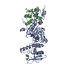
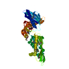
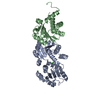
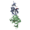
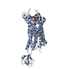
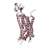
- PDB-4xnw: The human P2Y1 receptor in complex with MRS2500 -

+
Open data


ID or keywords:

Loading...

-
Basic information

Entry
Database: PDB / ID: 4xnw
TitleThe human P2Y1 receptor in complex with MRS2500
ComponentsP2Y purinoceptor 1,Rubredoxin,P2Y purinoceptor 1
KeywordsTRANSPORT PROTEIN / human P2Y1 receptor / G protein coupled receptor / platelet activation / membrane protein / PSI-Biology / Structural Genomics / GPCR Network / GPCR
Function / homology
Function and homology information


G protein-coupled ATP receptor activity / relaxation of muscle / A1 adenosine receptor binding / G protein-coupled ADP receptor activity / P2Y receptors / cellular response to purine-containing compound / positive regulation of inositol trisphosphate biosynthetic process / G protein-coupled purinergic nucleotide receptor activity / positive regulation of monoatomic ion transport / positive regulation of penile erection ...G protein-coupled ATP receptor activity / relaxation of muscle / A1 adenosine receptor binding / G protein-coupled ADP receptor activity / P2Y receptors / cellular response to purine-containing compound / positive regulation of inositol trisphosphate biosynthetic process / G protein-coupled purinergic nucleotide receptor activity / positive regulation of monoatomic ion transport / positive regulation of penile erection / negative regulation of norepinephrine secretion / glial cell migration / regulation of presynaptic cytosolic calcium ion concentration / G protein-coupled adenosine receptor signaling pathway / signal transduction involved in regulation of gene expression / positive regulation of hormone secretion / response to growth factor / cellular response to ATP / regulation of synaptic vesicle exocytosis / eating behavior / monoatomic ion transport / response to mechanical stimulus / presynaptic active zone membrane / adenylate cyclase-inhibiting G protein-coupled receptor signaling pathway / blood vessel diameter maintenance / establishment of localization in cell / ADP binding / protein localization to plasma membrane / cilium / platelet activation / ADP signalling through P2Y purinoceptor 1 / signaling receptor activity / phospholipase C-activating G protein-coupled receptor signaling pathway / cell body / positive regulation of cytosolic calcium ion concentration / regulation of cell shape / postsynaptic membrane / scaffold protein binding / G alpha (q) signalling events / basolateral plasma membrane / postsynaptic density / electron transfer activity / positive regulation of ERK1 and ERK2 cascade / cell surface receptor signaling pathway / G protein-coupled receptor signaling pathway / apical plasma membrane / iron ion binding / positive regulation of protein phosphorylation / protein heterodimerization activity / dendrite / glutamatergic synapse / cell surface / positive regulation of transcription by RNA polymerase II / ATP binding / plasma membrane
Similarity search - Function
P2Y purinoceptor 1 / Rubredoxin / Rubredoxin, iron-binding site / Rubredoxin signature. / Rubredoxin domain / Rubredoxin / Rubredoxin-like domain / Rubredoxin-like domain profile. / G-protein coupled receptors family 1 signature. / G protein-coupled receptor, rhodopsin-like ...P2Y purinoceptor 1 / Rubredoxin / Rubredoxin, iron-binding site / Rubredoxin signature. / Rubredoxin domain / Rubredoxin / Rubredoxin-like domain / Rubredoxin-like domain profile. / G-protein coupled receptors family 1 signature. / G protein-coupled receptor, rhodopsin-like / GPCR, rhodopsin-like, 7TM / G-protein coupled receptors family 1 profile. / 7 transmembrane receptor (rhodopsin family)
Similarity search - Domain/homology
Chem-2ID / Rubredoxin / P2Y purinoceptor 1
Similarity search - Component
Biological speciesHomo sapiens (human)
Clostridium pasteurianum (bacteria)
MethodX-RAY DIFFRACTION / SYNCHROTRON / Resolution: 2.7 Å
AuthorsZhang, D. / Gao, Z. / Jacobson, K. / Han, G.W. / Stevens, R. / Zhao, Q. / Wu, B. / GPCR Network (GPCR)
CitationJournal: Nature / Year: 2015
Title: Two disparate ligand-binding sites in the human P2Y1 receptor
Authors: Zhang, D. / Gao, Z.G. / Zhang, K. / Kiselev, E. / Crane, S. / Wang, J. / Paoletta, S. / Yi, C. / Ma, L. / Zhang, W. / Han, G.W. / Liu, H. / Cherezov, V. / Katritch, V. / Jiang, H. / Stevens, ...Authors: Zhang, D. / Gao, Z.G. / Zhang, K. / Kiselev, E. / Crane, S. / Wang, J. / Paoletta, S. / Yi, C. / Ma, L. / Zhang, W. / Han, G.W. / Liu, H. / Cherezov, V. / Katritch, V. / Jiang, H. / Stevens, R.C. / Jacobson, K.A. / Zhao, Q. / Wu, B.
History
DepositionJan 16, 2015Deposition site: RCSB / Processing site: PDBJ
Revision 1.0Apr 1, 2015Provider: repository / Type: Initial release
Revision 1.1Apr 22, 2015Group: Database references
Revision 1.2Apr 29, 2015Group: Structure summary
Revision 1.3Feb 5, 2020Group: Data collection / Database references ...Data collection / Database references / Derived calculations / Source and taxonomy / Structure summary
Category: audit_author / citation ...audit_author / citation / diffrn_source / entity_src_gen / pdbx_struct_oper_list / struct_keywords
Item: _citation.journal_id_CSD / _diffrn_source.pdbx_synchrotron_site ..._citation.journal_id_CSD / _diffrn_source.pdbx_synchrotron_site / _entity_src_gen.pdbx_alt_source_flag / _pdbx_struct_oper_list.symmetry_operation / _struct_keywords.text

-
Structure visualization

Structure viewerMolecule:
MolmilJmol/JSmol

Downloads & links

-
Assembly

Deposited unit
A: P2Y purinoceptor 1,Rubredoxin,P2Y purinoceptor 1
C: P2Y purinoceptor 1,Rubredoxin,P2Y purinoceptor 1
hetero molecules


Theoretical massNumber of molelcules
Total (without water)96,1906
Polymers94,9372
Non-polymers1,2534
Water18010
1
A: P2Y purinoceptor 1,Rubredoxin,P2Y purinoceptor 1
hetero molecules


Theoretical massNumber of molelcules
Total (without water)48,0953
Polymers47,4681
Non-polymers6272
Water181
TypeNameSymmetry operationNumber
identity operation1_555x,y,z1
2
C: P2Y purinoceptor 1,Rubredoxin,P2Y purinoceptor 1
hetero molecules


Theoretical massNumber of molelcules
Total (without water)48,0953
Polymers47,4681
Non-polymers6272
Water181
TypeNameSymmetry operationNumber
identity operation1_555x,y,z1
Unit cell
Length a, b, c (Å)40.670, 64.970, 77.570
Angle α, β, γ (deg.)101.82, 95.70, 100.85
Int Tables number1
Space group name H-MP1
Noncrystallographic symmetry (NCS)NCS domain:
IDEns-IDDetails
11A
21C
12A
22C

NCS domain segments:
Dom-IDComponent-IDEns-IDRefine codeAuth asym-IDAuth seq-ID
1010A38 - 333
2010C38 - 333
1020A1001 - 1054
2020C1001 - 1054

NCS ensembles :
ID
1
2

-
Components

#1: Protein P2Y purinoceptor 1,Rubredoxin,P2Y purinoceptor 1 / P2Y1 / ATP receptor / Purinergic receptor / Rd


Mass: 47468.289 Da / Num. of mol.: 2 / Mutation: D320N
Source method: isolated from a genetically manipulated source
Details: Chimera protein of P2Y purinoceptor 1 (P2RY1_HUMA) with Rubredoxin (RUBR_CLOPA) inserted into ICL3 domain between residues 247LYS and 253PRO, and replaced residues 248ASN to 252SER.
Source: (gene. exp.) Homo sapiens (human), (gene. exp.) Clostridium pasteurianum (bacteria)
Gene: P2RY1 / Production host: Spodoptera (butterflies/moths) / References: UniProt: P47900, UniProt: P00268
#2: Chemical ChemComp-2ID / [(1R,2S,4S,5S)-4-[2-iodo-6-(methylamino)-9H-purin-9-yl]-2-(phosphonooxy)bicyclo[3.1.0]hex-1-yl]methyl dihydrogen phosphate


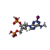
Mass: 561.163 Da / Num. of mol.: 2 / Source method: obtained synthetically / Formula: C13H18IN5O8P2
#3: Chemical ChemComp-ZN / ZINC ION


Mass: 65.409 Da / Num. of mol.: 2 / Source method: obtained synthetically / Formula: Zn
#4: Water ChemComp-HOH / water / Water


Mass: 18.015 Da / Num. of mol.: 10 / Source method: isolated from a natural source / Formula: H2O

-
Experimental details

-
Experiment

ExperimentMethod: X-RAY DIFFRACTION / Number of used crystals: 1

-
Sample preparation

CrystalDensity Matthews: 2.02 Å3/Da / Density % sol: 39.24 %
Crystal growTemperature: 293 K / Method: lipidic cubic phase / Details: PEG400 Sodium citrate HEPES

-
Data collection

DiffractionMean temperature: 100 K
Diffraction sourceSource: SYNCHROTRON / Site: SPring-8 / Beamline: BL41XU / Wavelength: 1 Å
DetectorType: MARMOSAIC 225 mm CCD / Detector: CCD / Date: Dec 8, 2013
RadiationProtocol: SINGLE WAVELENGTH / Monochromatic (M) / Laue (L): M / Scattering type: x-ray
Radiation wavelengthWavelength: 1 Å / Relative weight: 1
ReflectionResolution: 2.7→50 Å / Num. obs: 20471 / % possible obs: 98.2 % / Redundancy: 4.9 % / Net I/σ(I): 18.9

-
Processing

Software
NameVersionClassification
REFMAC5.7.0029refinement
HKL-2000data reduction
SCALEPACKdata scaling
PHASERphasing
RefinementResolution: 2.7→29.31 Å / Cor.coef. Fo:Fc: 0.924 / Cor.coef. Fo:Fc free: 0.896 / SU B: 8.27 / SU ML: 0.184 / Cross valid method: THROUGHOUT / ESU R Free: 0.085 / Stereochemistry target values: MAXIMUM LIKELIHOOD / Details: HYDROGENS HAVE BEEN ADDED IN THE RIDING POSITIONS
RfactorNum. reflection% reflectionSelection details
Rfree0.26714 1027 5.1 %RANDOM
Rwork0.21834 ---
obs0.22094 19283 98.1 %-
Solvent computationIon probe radii: 0.8 Å / Shrinkage radii: 0.8 Å / VDW probe radii: 1.2 Å / Solvent model: MASK
Displacement parametersBiso mean: 48.213 Å2
Baniso -1Baniso -2Baniso -3
1-13.03 Å24.64 Å20.66 Å2
2--8.83 Å2-3.55 Å2
3----21.86 Å2
Refinement stepCycle: LAST / Resolution: 2.7→29.31 Å
ProteinNucleic acidLigandSolventTotal
Num. atoms5569 0 60 10 5639
Refine LS restraints
Refine-IDTypeDev idealDev ideal targetNumber
X-RAY DIFFRACTIONr_bond_refined_d0.0090.025794
X-RAY DIFFRACTIONr_bond_other_d0.0040.025520
X-RAY DIFFRACTIONr_angle_refined_deg1.3311.9737902
X-RAY DIFFRACTIONr_angle_other_deg0.9873.00312641
X-RAY DIFFRACTIONr_dihedral_angle_1_deg5.335694
X-RAY DIFFRACTIONr_dihedral_angle_2_deg34.36222.345226
X-RAY DIFFRACTIONr_dihedral_angle_3_deg16.58715928
X-RAY DIFFRACTIONr_dihedral_angle_4_deg17.5331527
X-RAY DIFFRACTIONr_chiral_restr0.0760.2901
X-RAY DIFFRACTIONr_gen_planes_refined0.0060.0216342
X-RAY DIFFRACTIONr_gen_planes_other0.0030.021395
X-RAY DIFFRACTIONr_nbd_refined
X-RAY DIFFRACTIONr_nbd_other
X-RAY DIFFRACTIONr_nbtor_refined
X-RAY DIFFRACTIONr_nbtor_other
X-RAY DIFFRACTIONr_xyhbond_nbd_refined
X-RAY DIFFRACTIONr_xyhbond_nbd_other
X-RAY DIFFRACTIONr_metal_ion_refined
X-RAY DIFFRACTIONr_metal_ion_other
X-RAY DIFFRACTIONr_symmetry_vdw_refined
X-RAY DIFFRACTIONr_symmetry_vdw_other
X-RAY DIFFRACTIONr_symmetry_hbond_refined
X-RAY DIFFRACTIONr_symmetry_hbond_other
X-RAY DIFFRACTIONr_symmetry_metal_ion_refined
X-RAY DIFFRACTIONr_symmetry_metal_ion_other
X-RAY DIFFRACTIONr_mcbond_it
X-RAY DIFFRACTIONr_mcbond_other
X-RAY DIFFRACTIONr_mcangle_it
X-RAY DIFFRACTIONr_mcangle_other
X-RAY DIFFRACTIONr_scbond_it
X-RAY DIFFRACTIONr_scbond_other
X-RAY DIFFRACTIONr_scangle_it
X-RAY DIFFRACTIONr_scangle_other
X-RAY DIFFRACTIONr_long_range_B_refined
X-RAY DIFFRACTIONr_long_range_B_other
X-RAY DIFFRACTIONr_rigid_bond_restr
X-RAY DIFFRACTIONr_sphericity_free
X-RAY DIFFRACTIONr_sphericity_bonded
Refine LS restraints NCS

Refine-ID: X-RAY DIFFRACTION / Type: interatomic distance / Rms dev position: 0.12 Å / Weight position: 0.05

Ens-IDDom-IDAuth asym-IDNumber
11A16976
12C16976
21A2684
22C2684
LS refinement shellResolution: 2.7→2.769 Å / Total num. of bins used: 20
RfactorNum. reflection% reflection
Rfree0.381 71 -
Rwork0.303 1370 -
obs--97.36 %

+
About Yorodumi

-
News

-
Feb 9, 2022. New format data for meta-information of EMDB entries

New format data for meta-information of EMDB entries

  • Version 3 of the EMDB header file is now the official format.
  • The previous official version 1.9 will be removed from the archive.

Related info.:EMDB header

External links:wwPDB to switch to version 3 of the EMDB data model

-
Aug 12, 2020. Covid-19 info

Covid-19 info

URL: https://pdbj.org/emnavi/covid19.php

New page: Covid-19 featured information page in EM Navigator.

Related info.:Covid-19 info / Mar 5, 2020. Novel coronavirus structure data

+
Mar 5, 2020. Novel coronavirus structure data

Novel coronavirus structure data

Related info.:Yorodumi Speices / Aug 12, 2020. Covid-19 info

External links:COVID-19 featured content - PDBj / Molecule of the Month (242):Coronavirus Proteases

+
Jan 31, 2019. EMDB accession codes are about to change! (news from PDBe EMDB page)

EMDB accession codes are about to change! (news from PDBe EMDB page)

  • The allocation of 4 digits for EMDB accession codes will soon come to an end. Whilst these codes will remain in use, new EMDB accession codes will include an additional digit and will expand incrementally as the available range of codes is exhausted. The current 4-digit format prefixed with “EMD-” (i.e. EMD-XXXX) will advance to a 5-digit format (i.e. EMD-XXXXX), and so on. It is currently estimated that the 4-digit codes will be depleted around Spring 2019, at which point the 5-digit format will come into force.
  • The EM Navigator/Yorodumi systems omit the EMD- prefix.

Related info.:Q: What is EMD? / ID/Accession-code notation in Yorodumi/EM Navigator

External links:EMDB Accession Codes are Changing Soon! / Contact to PDBj

+
Jul 12, 2017. Major update of PDB

Major update of PDB

  • wwPDB released updated PDB data conforming to the new PDBx/mmCIF dictionary.
  • This is a major update changing the version number from 4 to 5, and with Remediation, in which all the entries are updated.
  • In this update, many items about electron microscopy experimental information are reorganized (e.g. em_software).
  • Now, EM Navigator and Yorodumi are based on the updated data.

External links:wwPDB Remediation / Enriched Model Files Conforming to OneDep Data Standards Now Available in the PDB FTP Archive

-
Yorodumi

Thousand views of thousand structures

  • Yorodumi is a browser for structure data from EMDB, PDB, SASBDB, etc.
  • This page is also the successor to EM Navigator detail page, and also detail information page/front-end page for Omokage search.
  • The word "yorodu" (or yorozu) is an old Japanese word meaning "ten thousand". "mi" (miru) is to see.

Related info.:EMDB / PDB / SASBDB / Comparison of 3 databanks / Yorodumi Search / Aug 31, 2016. New EM Navigator & Yorodumi / Yorodumi Papers / Jmol/JSmol / Function and homology information / Changes in new EM Navigator and Yorodumi

Read more