 Movie
Movie Controller

[English] 日本語
 Yorodumi
- PDB-4xi1: Crystal structure of U-box 2 of LubX / LegU2 / Lpp2887 from Legio... -
+ Open data

- Basic information
| Entry | Database: PDB / ID: 4xi1 | |||||||||
|---|---|---|---|---|---|---|---|---|---|---|
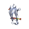
| Title | Crystal structure of U-box 2 of LubX / LegU2 / Lpp2887 from Legionella pneumophila str. Paris, wild-type | |||||||||
 Components Components | E3 ubiquitin-protein ligase LubX | |||||||||
 Keywords Keywords | LIGASE / ALPHA/BETA PROTEIN / EFFECTOR / STRUCTURAL GENOMICS / PSI-BIOLOGY / MIDWEST CENTER FOR STRUCTURAL GENOMICS / MCSG | |||||||||
| Function / homology |  Function and homology information Function and homology informationcellular response to misfolded protein / positive regulation of proteolysis / protein quality control for misfolded or incompletely synthesized proteins / RING-type E3 ubiquitin transferase / protein polyubiquitination / ubiquitin-protein transferase activity / ubiquitin protein ligase activity / host cell / protein-folding chaperone binding / proteasome-mediated ubiquitin-dependent protein catabolic process ...cellular response to misfolded protein / positive regulation of proteolysis / protein quality control for misfolded or incompletely synthesized proteins / RING-type E3 ubiquitin transferase / protein polyubiquitination / ubiquitin-protein transferase activity / ubiquitin protein ligase activity / host cell / protein-folding chaperone binding / proteasome-mediated ubiquitin-dependent protein catabolic process / protein ubiquitination / extracellular region / cytoplasm Similarity search - Function | |||||||||
| Biological species |   Legionella pneumophila (bacteria) Legionella pneumophila (bacteria) | |||||||||
| Method |  X-RAY DIFFRACTION / SYNCHROTRON / MOLECULAR REPLACEMENT / Resolution: 2.983 Å X-RAY DIFFRACTION / SYNCHROTRON / MOLECULAR REPLACEMENT / Resolution: 2.983 Å | |||||||||
 Authors Authors | Stogios, P.J. / Quaile, T. / Skarina, T. / Cuff, M. / Di Leo, R. / Yim, V. / Savchenko, A. / Joachimiak, A. / Midwest Center for Structural Genomics (MCSG) | |||||||||
 Citation Citation | Journal: Structure / Year: 2015 Title: Molecular Characterization of LubX: Functional Divergence of the U-Box Fold by Legionella pneumophila. Authors: Quaile, A.T. / Urbanus, M.L. / Stogios, P.J. / Nocek, B. / Skarina, T. / Ensminger, A.W. / Savchenko, A. | |||||||||
| History |
|
- Structure visualization
| Structure viewer | Molecule: MolmilJmol/JSmol |
|---|
- Downloads & links
-Download
| PDBx/mmCIF format | 4xi1.cif.gz | 112.2 KB | Display | PDBx/mmCIF format |
|---|---|---|---|---|
| PDB format | pdb4xi1.ent.gz | 88.2 KB | Display | PDB format |
| PDBx/mmJSON format | 4xi1.json.gz | Tree view | PDBx/mmJSON format | |
| Others |  Other downloads Other downloads |
-Validation report
| Arichive directory | https://data.pdbj.org/pub/pdb/validation_reports/xi/4xi1ftp://data.pdbj.org/pub/pdb/validation_reports/xi/4xi1 | HTTPS FTP |
|---|
-Related structure data
| Related structure data |  4wz0C  4wz2SC  4wz3C C: citing same article ( S: Starting model for refinement |
|---|---|
| Similar structure data | |
| Other databases |


-Links
 PDBj
PDBj
- Assembly
| Deposited unit | 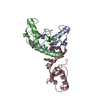
| ||||||||||||
|---|---|---|---|---|---|---|---|---|---|---|---|---|---|
| 1 | 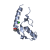
| ||||||||||||
| 2 | 
| ||||||||||||
| 3 | 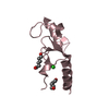
| ||||||||||||
| 4 | 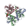
| ||||||||||||
| Unit cell |
| ||||||||||||
| Components on special symmetry positions |
|
-Components
| #1: Protein | Mass: 11774.493 Da / Num. of mol.: 3 / Fragment: UNP residues 102-202 Source method: isolated from a genetically manipulated source Source: (gene. exp.) Legionella pneumophila (strain Paris) (bacteria)Strain: Paris / Gene: lubX, lpp2887 / Plasmid: P15TV-LIC / Production host: References: UniProt: Q5X159, Ligases; Forming carbon-nitrogen bonds; Acid-amino-acid ligases (peptide synthases) #2: Chemical |   Mass: 35.453 Da / Num. of mol.: 2 / Source method: obtained synthetically / Formula: Cl Mass: 35.453 Da / Num. of mol.: 2 / Source method: obtained synthetically / Formula: Cl#3: Chemical | ChemComp-HEZ /   Mass: 118.174 Da / Num. of mol.: 5 / Source method: obtained synthetically / Formula: C6H14O2 Mass: 118.174 Da / Num. of mol.: 5 / Source method: obtained synthetically / Formula: C6H14O2#4: Chemical | ChemComp-GOL / |   Mass: 92.094 Da / Num. of mol.: 1 / Source method: obtained synthetically / Formula: C3H8O3 Mass: 92.094 Da / Num. of mol.: 1 / Source method: obtained synthetically / Formula: C3H8O3#5: Water | ChemComp-HOH / |  Mass: 18.015 Da / Num. of mol.: 87 / Source method: isolated from a natural source / Formula: H2O Mass: 18.015 Da / Num. of mol.: 87 / Source method: isolated from a natural source / Formula: H2O |
|---|
-Experimental details
-Experiment
| Experiment | Method: X-RAY DIFFRACTION / Number of used crystals: 1 |
|---|
- Sample preparation
| Crystal | Density Matthews: 4.85 Å3/Da / Density % sol: 74.65 % |
|---|---|
| Crystal grow | Temperature: 298 K / Method: vapor diffusion, hanging drop / pH: 7.5 Details: 15 mg/ml protein, V8 protease (1:100), 1.6 M ammonium sulfate, 0.1 M HEPES pH 7.5 and 2% hexanediol |
-Data collection
| Diffraction |
| |||||||||
|---|---|---|---|---|---|---|---|---|---|---|
| Diffraction source | Source: SYNCHROTRON / Site: APS  / Beamline: 19-ID / Wavelength: 0.9790433 Å / Beamline: 19-ID / Wavelength: 0.9790433 Å | |||||||||
| Detector | Type: ADSC QUANTUM 315 / Detector: CCD / Date: Dec 7, 2012 | |||||||||
| Radiation | Protocol: SINGLE WAVELENGTH / Monochromatic (M) / Laue (L): M / Scattering type: x-ray | |||||||||
| Radiation wavelength | Wavelength: 0.9790433 Å / Relative weight: 1 | |||||||||
| Reflection | Resolution: 2.98→20 Å / Num. obs: 13171 / % possible obs: 88.7 % / Redundancy: 5.7 % / Rmerge(I) obs: 0.08 / Net I/σ(I): 23.58 | |||||||||
| Reflection shell | Resolution: 2.98→3.03 Å / Redundancy: 5.6 % / Rmerge(I) obs: 0.623 / Mean I/σ(I) obs: 2.7 / % possible all: 92.7 |
- Processing
| Software |
| ||||||||||||||||||||||||||||||||||||||||||||||||||||||||||||||||||||||||||||||||||||||||||||||||||||||||||||||||||||||||||||||||||||||||||||||||||||||||||||||||||||||||||||||||||||||||||||||||||||||||||||||||||||||||||||||||||||||||||||||||||||||||||
|---|---|---|---|---|---|---|---|---|---|---|---|---|---|---|---|---|---|---|---|---|---|---|---|---|---|---|---|---|---|---|---|---|---|---|---|---|---|---|---|---|---|---|---|---|---|---|---|---|---|---|---|---|---|---|---|---|---|---|---|---|---|---|---|---|---|---|---|---|---|---|---|---|---|---|---|---|---|---|---|---|---|---|---|---|---|---|---|---|---|---|---|---|---|---|---|---|---|---|---|---|---|---|---|---|---|---|---|---|---|---|---|---|---|---|---|---|---|---|---|---|---|---|---|---|---|---|---|---|---|---|---|---|---|---|---|---|---|---|---|---|---|---|---|---|---|---|---|---|---|---|---|---|---|---|---|---|---|---|---|---|---|---|---|---|---|---|---|---|---|---|---|---|---|---|---|---|---|---|---|---|---|---|---|---|---|---|---|---|---|---|---|---|---|---|---|---|---|---|---|---|---|---|---|---|---|---|---|---|---|---|---|---|---|---|---|---|---|---|---|---|---|---|---|---|---|---|---|---|---|---|---|---|---|---|---|---|---|---|---|---|---|---|---|---|---|---|---|---|---|---|---|
| Refinement | Method to determine structure: MOLECULAR REPLACEMENT Starting model: 4WZ2 Resolution: 2.983→19.874 Å / SU ML: 0.35 / Cross valid method: FREE R-VALUE / σ(F): 1.35 / Phase error: 22.42 / Stereochemistry target values: ML
| ||||||||||||||||||||||||||||||||||||||||||||||||||||||||||||||||||||||||||||||||||||||||||||||||||||||||||||||||||||||||||||||||||||||||||||||||||||||||||||||||||||||||||||||||||||||||||||||||||||||||||||||||||||||||||||||||||||||||||||||||||||||||||
| Solvent computation | Shrinkage radii: 0.9 Å / VDW probe radii: 1.11 Å / Solvent model: FLAT BULK SOLVENT MODEL | ||||||||||||||||||||||||||||||||||||||||||||||||||||||||||||||||||||||||||||||||||||||||||||||||||||||||||||||||||||||||||||||||||||||||||||||||||||||||||||||||||||||||||||||||||||||||||||||||||||||||||||||||||||||||||||||||||||||||||||||||||||||||||
| Refinement step | Cycle: LAST / Resolution: 2.983→19.874 Å
| ||||||||||||||||||||||||||||||||||||||||||||||||||||||||||||||||||||||||||||||||||||||||||||||||||||||||||||||||||||||||||||||||||||||||||||||||||||||||||||||||||||||||||||||||||||||||||||||||||||||||||||||||||||||||||||||||||||||||||||||||||||||||||
| Refine LS restraints |
| ||||||||||||||||||||||||||||||||||||||||||||||||||||||||||||||||||||||||||||||||||||||||||||||||||||||||||||||||||||||||||||||||||||||||||||||||||||||||||||||||||||||||||||||||||||||||||||||||||||||||||||||||||||||||||||||||||||||||||||||||||||||||||
| LS refinement shell |
| ||||||||||||||||||||||||||||||||||||||||||||||||||||||||||||||||||||||||||||||||||||||||||||||||||||||||||||||||||||||||||||||||||||||||||||||||||||||||||||||||||||||||||||||||||||||||||||||||||||||||||||||||||||||||||||||||||||||||||||||||||||||||||
| Refinement TLS params. | Method: refined / Refine-ID: X-RAY DIFFRACTION
| ||||||||||||||||||||||||||||||||||||||||||||||||||||||||||||||||||||||||||||||||||||||||||||||||||||||||||||||||||||||||||||||||||||||||||||||||||||||||||||||||||||||||||||||||||||||||||||||||||||||||||||||||||||||||||||||||||||||||||||||||||||||||||
| Refinement TLS group |
|
