 Movie
Movie Controller


+ Open data

- Basic information
| Entry | Database: PDB / ID: 4xd7 | ||||||
|---|---|---|---|---|---|---|---|
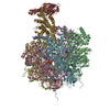
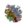
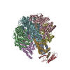
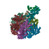
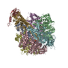
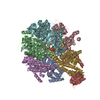
| Title | Structure of thermophilic F1-ATPase inhibited by epsilon subunit | ||||||
 Components Components | (ATP synthase ...) x 4 | ||||||
 Keywords Keywords | HYDROLASE / F1-ATPASE / ATP SYNTHASE / ROTARY MOTOR PROTEIN / ROTATIONAL CATALYSIS / BACILLUS PS3 / THERMOPHILIC | ||||||
| Function / homology |  Function and homology information Function and homology information: / proton motive force-driven plasma membrane ATP synthesis / proton-transporting ATPase activity, rotational mechanism / H+-transporting two-sector ATPase / proton-transporting ATP synthase complex / proton-transporting ATP synthase activity, rotational mechanism / ADP binding / ATP hydrolysis activity / ATP binding / plasma membrane Similarity search - Function | ||||||
| Biological species |  | ||||||
| Method |  X-RAY DIFFRACTION / SYNCHROTRON / Resolution: 3.9 Å X-RAY DIFFRACTION / SYNCHROTRON / Resolution: 3.9 Å | ||||||
 Authors Authors | SHIRAKIHARA, Y. / SHIRATORI, A. / TANIKAWA, H. / NAKASAKO, M. / YOSHIDA, M. / SUZUKI, T. | ||||||
 Citation Citation | Journal: Febs J. / Year: 2015 Title: Structure of a thermophilic F1 -ATPase inhibited by an epsilon-subunit: deeper insight into the epsilon-inhibition mechanism. Authors: Shirakihara, Y. / Shiratori, A. / Tanikawa, H. / Nakasako, M. / Yoshida, M. / Suzuki, T. | ||||||
| History |
|
- Structure visualization
| Structure viewer | Molecule: MolmilJmol/JSmol |
|---|
- Downloads & links
-Download
| PDBx/mmCIF format | 4xd7.cif.gz | 589 KB | Display | PDBx/mmCIF format |
|---|---|---|---|---|
| PDB format | pdb4xd7.ent.gz | 471.8 KB | Display | PDB format |
| PDBx/mmJSON format | 4xd7.json.gz | Tree view | PDBx/mmJSON format | |
| Others |  Other downloads Other downloads |
-Validation report
| Arichive directory | https://data.pdbj.org/pub/pdb/validation_reports/xd/4xd7ftp://data.pdbj.org/pub/pdb/validation_reports/xd/4xd7 | HTTPS FTP |
|---|
-Related structure data


-Links
 PDBj
PDBj


- Assembly
| Deposited unit | 
| ||||||||
|---|---|---|---|---|---|---|---|---|---|
| 1 |
| ||||||||
| Unit cell |
|
-Components
-ATP synthase ... , 4 types, 8 molecules ABCDEFGH
| #1: Protein | Mass: 55317.543 Da / Num. of mol.: 3 Source method: isolated from a genetically manipulated source Source: (gene. exp.) References: UniProt: Q5KUJ1, UniProt: P09219*PLUS, H+-transporting two-sector ATPase #2: Protein | Mass: 53949.949 Da / Num. of mol.: 3 Source method: isolated from a genetically manipulated source Source: (gene. exp.) References: UniProt: Q5KUJ3, UniProt: P07677*PLUS, H+-transporting two-sector ATPase #3: Protein | | Mass: 32297.641 Da / Num. of mol.: 1 Source method: isolated from a genetically manipulated source Source: (gene. exp.) #4: Protein | | Mass: 14773.485 Da / Num. of mol.: 1 Source method: isolated from a genetically manipulated source Source: (gene. exp.) |
|---|
-Non-polymers , 2 types, 5 molecules 

| #5: Chemical | ChemComp-SO4 /  Mass: 96.063 Da / Num. of mol.: 4 / Source method: obtained synthetically / Formula: SO4 Mass: 96.063 Da / Num. of mol.: 4 / Source method: obtained synthetically / Formula: SO4#6: Chemical | ChemComp-ADP / |  Mass: 427.201 Da / Num. of mol.: 1 / Source method: obtained synthetically / Formula: C10H15N5O10P2 / Comment: ADP, energy-carrying molecule*YM Mass: 427.201 Da / Num. of mol.: 1 / Source method: obtained synthetically / Formula: C10H15N5O10P2 / Comment: ADP, energy-carrying molecule*YM |
|---|
-Details
| Has protein modification | Y |
|---|---|
| Sequence details | Author states that the sequence reported for this entry (Bacillus PS3 F1) matches better to that of ...Author states that the sequence reported for this entry (Bacillus PS3 F1) matches better to that of available UNP reference sequence for Geobacillus kaustophilus (strain HTA426) 235909, so Q5KUJ1, Q5KUJ2, Q5KUJ3 and Q5KUJ4 were selected as database reference sequence in this structure instead of UNP P09219 ,P07677,P09222 and P07678 which are outdated. The accession number at GenBank are LC076382 (Bacillus_PS3_alpha, Bacillus_PS3_gamma, Bacillus_PS3_beta) and LC076383 (Bacillus_PS3_ epsilon). |
-Experimental details
-Experiment
| Experiment | Method: X-RAY DIFFRACTION / Number of used crystals: 1 |
|---|
- Sample preparation
| Crystal | Density Matthews: 2.79 Å3/Da / Density % sol: 55.93 % |
|---|---|
| Crystal grow | Temperature: 298 K / Method: vapor diffusion, sitting drop Details: [drop]10 % PEG 6,000, 0.20 M sodium chloride, 0.05 M Tris-sulphate buffer (pH 8.0), 2mM DTT, 5mM CyDTA, 10% (v/v) ethyleneglycol and 10 mg/ml protein/ [reservoir] 14-16 % PEG 6,000, 0.2 M ...Details: [drop]10 % PEG 6,000, 0.20 M sodium chloride, 0.05 M Tris-sulphate buffer (pH 8.0), 2mM DTT, 5mM CyDTA, 10% (v/v) ethyleneglycol and 10 mg/ml protein/ [reservoir] 14-16 % PEG 6,000, 0.2 M sodium chloride, 0.05 M Tris-sulphate buffer (pH 8.0), 5mM CyDTA and 10% (v/v) ethyleneglycol |
-Data collection
| Diffraction | Mean temperature: 100 K |
|---|---|
| Diffraction source | Source: SYNCHROTRON / Site: Photon Factory  / Beamline: BL-6A / Wavelength: 0.97879 Å / Beamline: BL-6A / Wavelength: 0.97879 Å |
| Detector | Type: ADSC QUANTUM 315 / Detector: CCD / Date: Nov 22, 2007 |
| Radiation | Protocol: SINGLE WAVELENGTH / Monochromatic (M) / Laue (L): M / Scattering type: x-ray |
| Radiation wavelength | Wavelength: 0.97879 Å / Relative weight: 1 |
| Reflection | Resolution: 3.9→69.2 Å / Num. obs: 38420 / % possible obs: 99.6 % / Redundancy: 6.6 % / Rmerge(I) obs: 0.1 / Net I/σ(I): 14.7 |
| Reflection shell | Resolution: 3.9→4.11 Å / Redundancy: 5.7 % / Rmerge(I) obs: 0.52 / Mean I/σ(I) obs: 3.3 / % possible all: 97.9 |
- Processing
| Software |
| |||||||||||||||||||||||||||||||||||||||||||||||||||||||||||||||||||||||||||||||||||||||||||||||||||||||||||||||||||||||||||||||||||||||||||||||||||||||||||||||||||||||||||||||||||||||||||||
|---|---|---|---|---|---|---|---|---|---|---|---|---|---|---|---|---|---|---|---|---|---|---|---|---|---|---|---|---|---|---|---|---|---|---|---|---|---|---|---|---|---|---|---|---|---|---|---|---|---|---|---|---|---|---|---|---|---|---|---|---|---|---|---|---|---|---|---|---|---|---|---|---|---|---|---|---|---|---|---|---|---|---|---|---|---|---|---|---|---|---|---|---|---|---|---|---|---|---|---|---|---|---|---|---|---|---|---|---|---|---|---|---|---|---|---|---|---|---|---|---|---|---|---|---|---|---|---|---|---|---|---|---|---|---|---|---|---|---|---|---|---|---|---|---|---|---|---|---|---|---|---|---|---|---|---|---|---|---|---|---|---|---|---|---|---|---|---|---|---|---|---|---|---|---|---|---|---|---|---|---|---|---|---|---|---|---|---|---|---|---|
| Refinement | Starting model: 1SKY,1E79,1FS0 Resolution: 3.9→19.989 Å / SU ML: 0.43 / σ(F): 1.9 / Phase error: 27.65 / Stereochemistry target values: ML
| |||||||||||||||||||||||||||||||||||||||||||||||||||||||||||||||||||||||||||||||||||||||||||||||||||||||||||||||||||||||||||||||||||||||||||||||||||||||||||||||||||||||||||||||||||||||||||||
| Solvent computation | Shrinkage radii: 0.27 Å / VDW probe radii: 0.6 Å / Solvent model: FLAT BULK SOLVENT MODEL / Bsol: 88.442 Å2 / ksol: 0.31 e/Å3 | |||||||||||||||||||||||||||||||||||||||||||||||||||||||||||||||||||||||||||||||||||||||||||||||||||||||||||||||||||||||||||||||||||||||||||||||||||||||||||||||||||||||||||||||||||||||||||||
| Displacement parameters |
| |||||||||||||||||||||||||||||||||||||||||||||||||||||||||||||||||||||||||||||||||||||||||||||||||||||||||||||||||||||||||||||||||||||||||||||||||||||||||||||||||||||||||||||||||||||||||||||
| Refinement step | Cycle: LAST / Resolution: 3.9→19.989 Å
| |||||||||||||||||||||||||||||||||||||||||||||||||||||||||||||||||||||||||||||||||||||||||||||||||||||||||||||||||||||||||||||||||||||||||||||||||||||||||||||||||||||||||||||||||||||||||||||
| Refine LS restraints |
| |||||||||||||||||||||||||||||||||||||||||||||||||||||||||||||||||||||||||||||||||||||||||||||||||||||||||||||||||||||||||||||||||||||||||||||||||||||||||||||||||||||||||||||||||||||||||||||
| LS refinement shell |
|
