[English] 日本語
 Yorodumi
- PDB-1sky: CRYSTAL STRUCTURE OF THE NUCLEOTIDE FREE ALPHA3BETA3 SUB-COMPLEX ... -
+ Open data
- Basic information
| Entry | Database: PDB / ID: 1sky | ||||||
|---|---|---|---|---|---|---|---|
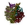
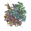
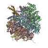
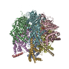
| Title | CRYSTAL STRUCTURE OF THE NUCLEOTIDE FREE ALPHA3BETA3 SUB-COMPLEX OF F1-ATPASE FROM THE THERMOPHILIC BACILLUS PS3 | ||||||
 Components Components | (F1-ATPASE) x 2 | ||||||
 Keywords Keywords | ATP SYNTHASE / F1FO ATP SYNTHASE / F1-ATPASE / ALPHA3BETA3 SUBCOMPLEX OF F1-ATPASE / HYDROLASE | ||||||
| Function / homology |  Function and homology information Function and homology informationH+-transporting two-sector ATPase / proton-transporting ATP synthase complex / proton-transporting ATP synthase activity, rotational mechanism / ADP binding / ATP hydrolysis activity / ATP binding / plasma membrane Similarity search - Function | ||||||
| Biological species |  | ||||||
| Method |  X-RAY DIFFRACTION / X-RAY DIFFRACTION /  SYNCHROTRON / SYNCHROTRON /  MOLECULAR REPLACEMENT / Resolution: 3.2 Å MOLECULAR REPLACEMENT / Resolution: 3.2 Å | ||||||
 Authors Authors | Shirakihara, Y. / Leslie, A.G.W. / Abrahams, J.P. / Walker, J.E. / Ueda, T. / Sekimoto, Y. / Kambara, M. / Saika, K. / Kagawa, Y. / Yoshida, M. | ||||||
 Citation Citation |  Journal: Structure / Year: 1997 Journal: Structure / Year: 1997Title: The crystal structure of the nucleotide-free alpha 3 beta 3 subcomplex of F1-ATPase from the thermophilic Bacillus PS3 is a symmetric trimer. Authors: Shirakihara, Y. / Leslie, A.G. / Abrahams, J.P. / Walker, J.E. / Ueda, T. / Sekimoto, Y. / Kambara, M. / Saika, K. / Kagawa, Y. / Yoshida, M. #1:  Journal: Photon Factory Activity Report / Year: 1994 Journal: Photon Factory Activity Report / Year: 1994Title: X-Ray Crystal Analysis of Alpha3Beta3 Complex of F1-ATPase from a Thermophilic Bacterium Ps3 Authors: Shirakihara, Y. / Ueda, T. / Sekimoto, Y. / Yoshida, M. / Saika, K. | ||||||
| History |
|
- Structure visualization
| Structure viewer | Molecule:  Molmil Molmil Jmol/JSmol Jmol/JSmol |
|---|
- Downloads & links
- Download
| PDBx/mmCIF format |  1sky.cif.gz 1sky.cif.gz | 190.8 KB | Display |  PDBx/mmCIF format PDBx/mmCIF format |
|---|---|---|---|---|
| PDB format |  pdb1sky.ent.gz pdb1sky.ent.gz | 151.2 KB | Display |  PDB format PDB format |
| PDBx/mmJSON format |  1sky.json.gz 1sky.json.gz | Tree view |  PDBx/mmJSON format PDBx/mmJSON format | |
| Others |  Other downloads Other downloads |
-Validation report
| Summary document |  1sky_validation.pdf.gz 1sky_validation.pdf.gz | 444.7 KB | Display |  wwPDB validaton report wwPDB validaton report |
|---|---|---|---|---|
| Full document |  1sky_full_validation.pdf.gz 1sky_full_validation.pdf.gz | 483.8 KB | Display | |
| Data in XML |  1sky_validation.xml.gz 1sky_validation.xml.gz | 37.8 KB | Display | |
| Data in CIF |  1sky_validation.cif.gz 1sky_validation.cif.gz | 51 KB | Display | |
| Arichive directory |  https://data.pdbj.org/pub/pdb/validation_reports/sk/1sky https://data.pdbj.org/pub/pdb/validation_reports/sk/1sky ftp://data.pdbj.org/pub/pdb/validation_reports/sk/1sky ftp://data.pdbj.org/pub/pdb/validation_reports/sk/1sky | HTTPS FTP |
-Related structure data
| Similar structure data |
|---|
- Links
Links
- Assembly
| Deposited unit | 
| ||||||||
|---|---|---|---|---|---|---|---|---|---|
| 1 | 
| ||||||||
| Unit cell |
|
- Components
| #1: Protein | Mass: 54648.328 Da / Num. of mol.: 1 Source method: isolated from a genetically manipulated source Source: (gene. exp.)  Plasmid: PKK ALPHA FOR ALPHA SUBUNIT, PUC118BETA FOR BETA SUBUNIT Production host:  | ||
|---|---|---|---|
| #2: Protein | Mass: 51998.082 Da / Num. of mol.: 1 Source method: isolated from a genetically manipulated source Source: (gene. exp.)  Plasmid: PKK ALPHA FOR ALPHA SUBUNIT, PUC118BETA FOR BETA SUBUNIT Production host:  | ||
| #3: Chemical | | Compound details | ALPHA3 BETA3 SUBCOMPLEX OF F1-ATPASE COMPRISES THREE ALPHA AND THREE BETA SUBUNITS. BECAUSE THE ...ALPHA3 BETA3 SUBCOMPLEX | |
-Experimental details
-Experiment
| Experiment | Method:  X-RAY DIFFRACTION / Number of used crystals: 1 X-RAY DIFFRACTION / Number of used crystals: 1 |
|---|
- Sample preparation
| Crystal | Density Matthews: 3.2 Å3/Da / Density % sol: 61 % Description: J.P. ABRAHAMS, A.G.W. LESLIE, R. LUTTER, J.E. WALKER STRUCTURE AT 2.8A RESOLUTION OF F1-ATPASE FROM BOVINE HEART MITOCHONDRIA, NATURE 370,621-628, (1994) | ||||||||||||||||||||||||||||||||||||||||
|---|---|---|---|---|---|---|---|---|---|---|---|---|---|---|---|---|---|---|---|---|---|---|---|---|---|---|---|---|---|---|---|---|---|---|---|---|---|---|---|---|---|
| Crystal grow | Temperature: 288 K / Method: vapor diffusion, hanging drop / pH: 8 Details: HANGING DROP WITH 9-11% PEG20000, 0.12 M SODIUM SULFATE AT PH8.0, EQUILIBRATED AGAINST PROTEIN FREE RESERVOIR WITH HIGHER PEG SOLUTION BY 2%. KEPT AT 15DEG., vapor diffusion - hanging drop, temperature 288K | ||||||||||||||||||||||||||||||||||||||||
| Crystal grow | *PLUS Temperature: 15 ℃ / Method: vapor diffusion, hanging drop | ||||||||||||||||||||||||||||||||||||||||
| Components of the solutions | *PLUS
|
-Data collection
| Diffraction | Mean temperature: 293 K |
|---|---|
| Diffraction source | Source:  SYNCHROTRON / Site: SYNCHROTRON / Site:  Photon Factory Photon Factory  / Beamline: BL-18B / Wavelength: 1 / Beamline: BL-18B / Wavelength: 1 |
| Detector | Detector: IMAGE PLATE / Date: May 24, 1994 / Details: MIRROR + MONOCHROMATOR |
| Radiation | Monochromatic (M) / Laue (L): M / Scattering type: x-ray |
| Radiation wavelength | Wavelength: 1 Å / Relative weight: 1 |
| Reflection | Resolution: 3.2→50 Å / Num. obs: 109106 / % possible obs: 99 % / Redundancy: 4.6 % / Biso Wilson estimate: 83 Å2 / Rmerge(I) obs: 0.075 / Net I/σ(I): 8.9 |
| Reflection shell | Resolution: 3.2→3.37 Å / Redundancy: 3.9 % / Rmerge(I) obs: 0.44 / Mean I/σ(I) obs: 1.5 / % possible all: 98.1 |
| Reflection | *PLUS Num. obs: 22429 / Num. measured all: 109106 |
| Reflection shell | *PLUS % possible obs: 98.1 % |
- Processing
| Software |
| ||||||||||||||||||||||||||||||||||||||||||||||||||||||||||||
|---|---|---|---|---|---|---|---|---|---|---|---|---|---|---|---|---|---|---|---|---|---|---|---|---|---|---|---|---|---|---|---|---|---|---|---|---|---|---|---|---|---|---|---|---|---|---|---|---|---|---|---|---|---|---|---|---|---|---|---|---|---|
| Refinement | Method to determine structure:  MOLECULAR REPLACEMENT MOLECULAR REPLACEMENTStarting model: MITOCHODRIAL F1-ATPASE Resolution: 3.2→6 Å
| ||||||||||||||||||||||||||||||||||||||||||||||||||||||||||||
| Displacement parameters | Biso mean: 68 Å2 | ||||||||||||||||||||||||||||||||||||||||||||||||||||||||||||
| Refine analyze | Luzzati coordinate error obs: 0.4 Å | ||||||||||||||||||||||||||||||||||||||||||||||||||||||||||||
| Refinement step | Cycle: LAST / Resolution: 3.2→6 Å
| ||||||||||||||||||||||||||||||||||||||||||||||||||||||||||||
| Refine LS restraints |
| ||||||||||||||||||||||||||||||||||||||||||||||||||||||||||||
| LS refinement shell | Resolution: 3.2→3.32 Å / Total num. of bins used: 8
| ||||||||||||||||||||||||||||||||||||||||||||||||||||||||||||
| Software | *PLUS Name:  X-PLOR / Version: 3.1 / Classification: refinement X-PLOR / Version: 3.1 / Classification: refinement | ||||||||||||||||||||||||||||||||||||||||||||||||||||||||||||
| Refine LS restraints | *PLUS
| ||||||||||||||||||||||||||||||||||||||||||||||||||||||||||||
| LS refinement shell | *PLUS Rfactor Rwork: 0.37 |
 Movie
Movie Controller


 PDBj
PDBj


