 Movie
Movie Controller

[English] 日本語
 Yorodumi
- PDB-3a5c: Inter-subunit interaction and quaternary rearrangement defined by... -
+ Open data

- Basic information
| Entry | Database: PDB / ID: 3a5c | ||||||
|---|---|---|---|---|---|---|---|
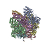
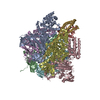
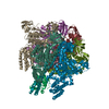
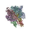
| Title | Inter-subunit interaction and quaternary rearrangement defined by the central stalk of prokaryotic V1-ATPase | ||||||
 Components Components |
| ||||||
 Keywords Keywords | HYDROLASE / V-ATPase / asymmetric / rotation / vacuolar type / ATP synthesis / ATP-binding / Hydrogen ion transport / Ion transport / Nucleotide-binding / Transport | ||||||
| Function / homology |  Function and homology information Function and homology informationproton motive force-driven plasma membrane ATP synthesis / proton-transporting ATPase activity, rotational mechanism / H+-transporting two-sector ATPase / proton-transporting ATP synthase complex / proton-transporting ATP synthase activity, rotational mechanism / ATP binding Similarity search - Function | ||||||
| Biological species |   Thermus thermophilus (bacteria) Thermus thermophilus (bacteria) | ||||||
| Method |  X-RAY DIFFRACTION / SYNCHROTRON / FOURIER SYNTHESIS / Resolution: 4.51 Å X-RAY DIFFRACTION / SYNCHROTRON / FOURIER SYNTHESIS / Resolution: 4.51 Å | ||||||
 Authors Authors | Numoto, N. / Hasegawa, Y. / Takeda, K. / Miki, K. | ||||||
 Citation Citation | Journal: Embo Rep. / Year: 2009 Title: Inter-subunit interaction and quaternary rearrangement defined by the central stalk of prokaryotic V1-ATPase Authors: Numoto, N. / Hasegawa, Y. / Takeda, K. / Miki, K. | ||||||
| History |
|
- Structure visualization
| Structure viewer | Molecule: MolmilJmol/JSmol |
|---|
- Downloads & links
-Download
| PDBx/mmCIF format | 3a5c.cif.gz | 859.7 KB | Display | PDBx/mmCIF format |
|---|---|---|---|---|
| PDB format | pdb3a5c.ent.gz | 582.1 KB | Display | PDB format |
| PDBx/mmJSON format | 3a5c.json.gz | Tree view | PDBx/mmJSON format | |
| Others |  Other downloads Other downloads |
-Validation report
| Arichive directory | https://data.pdbj.org/pub/pdb/validation_reports/a5/3a5cftp://data.pdbj.org/pub/pdb/validation_reports/a5/3a5c | HTTPS FTP |
|---|
-Related structure data
| Related structure data |  3a5dSC S: Starting model for refinement C: citing same article ( |
|---|---|
| Similar structure data |
-Links
 PDBj
PDBj


- Assembly
| Deposited unit | 
| ||||||||
|---|---|---|---|---|---|---|---|---|---|
| 1 | 
| ||||||||
| 2 | 
| ||||||||
| Unit cell |
|
-Components
| #1: Protein | Mass: 63699.980 Da / Num. of mol.: 6 / Source method: isolated from a natural source / Source: (natural) Thermus thermophilus (bacteria) / Strain: HB8References: UniProt: Q56403, H+-transporting two-sector ATPase #2: Protein | Mass: 53219.500 Da / Num. of mol.: 6 / Source method: isolated from a natural source / Source: (natural) Thermus thermophilus (bacteria) / Strain: HB8References: UniProt: Q56404, H+-transporting two-sector ATPase #3: Protein | Mass: 24715.566 Da / Num. of mol.: 2 / Source method: isolated from a natural source / Source: (natural) Thermus thermophilus (bacteria) / Strain: HB8References: UniProt: O87880, H+-transporting two-sector ATPase #4: Protein | Mass: 11294.904 Da / Num. of mol.: 2 / Source method: isolated from a natural source / Source: (natural) Thermus thermophilus (bacteria) / Strain: HB8References: UniProt: P74903, H+-transporting two-sector ATPase #5: Chemical | ChemComp-ADP /   Mass: 427.201 Da / Num. of mol.: 4 / Source method: obtained synthetically / Formula: C10H15N5O10P2 / Comment: ADP, energy-carrying molecule*YM Mass: 427.201 Da / Num. of mol.: 4 / Source method: obtained synthetically / Formula: C10H15N5O10P2 / Comment: ADP, energy-carrying molecule*YM |
|---|
-Experimental details
-Experiment
| Experiment | Method: X-RAY DIFFRACTION / Number of used crystals: 1 |
|---|
- Sample preparation
| Crystal | Density Matthews: 4.01 Å3/Da / Density % sol: 69.34 % |
|---|---|
| Crystal grow | Temperature: 293 K / Method: vapor diffusion, sitting drop / pH: 6 Details: 1.6M ammonium sulfate, 0.1M MES-NaOH (pH6.0), 10% dioxane, VAPOR DIFFUSION, SITTING DROP, temperature 293K |
-Data collection
| Diffraction | Mean temperature: 100 K |
|---|---|
| Diffraction source | Source: SYNCHROTRON / Site: SPring-8  / Beamline: BL41XU / Wavelength: 1 Å / Beamline: BL41XU / Wavelength: 1 Å |
| Detector | Date: Mar 5, 2009 |
| Radiation | Protocol: SINGLE WAVELENGTH / Monochromatic (M) / Laue (L): M / Scattering type: x-ray |
| Radiation wavelength | Wavelength: 1 Å / Relative weight: 1 |
| Reflection | Resolution: 4.5→50 Å / Num. obs: 70598 / % possible obs: 96.9 % / Redundancy: 5.1 % / Rsym value: 0.087 / Net I/σ(I): 10.3 |
| Reflection shell | Resolution: 4.5→4.66 Å / Redundancy: 2.8 % / Mean I/σ(I) obs: 2.3 / Num. unique all: 6683 / Rsym value: 0.417 / % possible all: 92.7 |
- Processing
| Software |
| ||||||||||||||||||||||||||||||||||||||||||||||||||||||||||||
|---|---|---|---|---|---|---|---|---|---|---|---|---|---|---|---|---|---|---|---|---|---|---|---|---|---|---|---|---|---|---|---|---|---|---|---|---|---|---|---|---|---|---|---|---|---|---|---|---|---|---|---|---|---|---|---|---|---|---|---|---|---|
| Refinement | Method to determine structure: FOURIER SYNTHESIS Starting model: PDB ENTRY 3A5D Resolution: 4.51→29.92 Å / Rfactor Rfree error: 0.007 / Data cutoff high absF: 11832712.93 / Data cutoff low absF: 0 / Isotropic thermal model: DOMAIN / Cross valid method: THROUGHOUT / σ(F): 0 Details: BULK SOLVENT MODEL USED, REFINEMENTS WERE PERFORMED BY THE RIGID BODY AND B-DOMAIN REFINEMENTS. THE DOMAIN DEFINITIONS WERE SET TO FOUR DOMAINS IN THE A SUBUNIT (AI: RESIDUES 1-70; AII: 71- ...Details: BULK SOLVENT MODEL USED, REFINEMENTS WERE PERFORMED BY THE RIGID BODY AND B-DOMAIN REFINEMENTS. THE DOMAIN DEFINITIONS WERE SET TO FOUR DOMAINS IN THE A SUBUNIT (AI: RESIDUES 1-70; AII: 71-198; AIII; 199-429; AIV: 430-577), THREE IN THE B SUBUNIT (BI: 7-80; BII: 81-359; BIII; 360-463), TWO IN THE D SUBUNIT (DI: 1-55 AND DII: 132-205), AND TWO REGIONS OF F (FI: 1-75 AND FII: 76-104), RESPECTIVELY.
| ||||||||||||||||||||||||||||||||||||||||||||||||||||||||||||
| Solvent computation | Solvent model: FLAT MODEL / Bsol: 160.757 Å2 / ksol: 0.1 e/Å3 | ||||||||||||||||||||||||||||||||||||||||||||||||||||||||||||
| Displacement parameters | Biso mean: 142.2 Å2
| ||||||||||||||||||||||||||||||||||||||||||||||||||||||||||||
| Refine analyze |
| ||||||||||||||||||||||||||||||||||||||||||||||||||||||||||||
| Refinement step | Cycle: LAST / Resolution: 4.51→29.92 Å
| ||||||||||||||||||||||||||||||||||||||||||||||||||||||||||||
| Refine LS restraints |
| ||||||||||||||||||||||||||||||||||||||||||||||||||||||||||||
| LS refinement shell | Resolution: 4.5→4.66 Å / Rfactor Rfree error: 0.028 / Total num. of bins used: 10
| ||||||||||||||||||||||||||||||||||||||||||||||||||||||||||||
| Xplor file |
|
