- PDB-3w3a: Crystal structure of V1-ATPase at 3.9 angstrom resolution -
+
Open data
ID or keywords:
Loading...
-
Basic information
Entry
Database: PDB / ID: 3w3a
Title
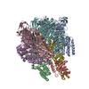
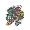
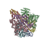
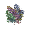
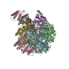
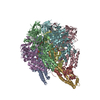
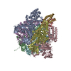
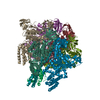
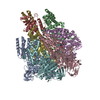
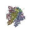
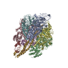
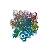
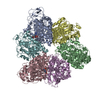
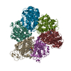
Crystal structure of V1-ATPase at 3.9 angstrom resolution
Components
V-type ATP synthase alpha chain
V-type ATP synthase beta chain
V-type ATP synthase subunit D
V-type ATP synthase subunit F
Keywords
HYDROLASE / ATP SYNTHESIS / HYDROGEN ION TRANSPORT / NUCLEOTIDE-BINDING / Catalytic Domain / Molecular Motor Proteins / Quaternary / Proton-Translocating ATPases / Thermus thermophilus / Vacuolar Proton-Translocating ATPases / Hydrolysis
Function / homology
Function and homology information
proton motive force-driven plasma membrane ATP synthesis / proton-transporting ATPase activity, rotational mechanism / H+-transporting two-sector ATPase / proton-transporting ATP synthase complex / proton-transporting ATP synthase activity, rotational mechanism / ATP binding Similarity search - Function
Helix Hairpins - #3240 / ATPase, V1 complex, subunit F / Rossmann fold - #12240 / ATPase, V1 complex, subunit F, bacterial/archaeal / Bovine Mitochondrial F1-ATPase, ATP Synthase Beta Chain; Chain D, domain3 / Bovine Mitochondrial F1-atpase; Atp Synthase Beta Chain; Chain D, domain 3 / ATPase, V1 complex, subunit D / ATPase, V1 complex, subunit F / ATPase, V1 complex, subunit F superfamily / ATP synthase subunit D ...Helix Hairpins - #3240 / ATPase, V1 complex, subunit F / Rossmann fold - #12240 / ATPase, V1 complex, subunit F, bacterial/archaeal / Bovine Mitochondrial F1-ATPase, ATP Synthase Beta Chain; Chain D, domain3 / Bovine Mitochondrial F1-atpase; Atp Synthase Beta Chain; Chain D, domain 3 / ATPase, V1 complex, subunit D / ATPase, V1 complex, subunit F / ATPase, V1 complex, subunit F superfamily / ATP synthase subunit D / ATP synthase (F/14-kDa) subunit / V-type ATP synthase regulatory subunit B/beta / V-type ATP synthase catalytic alpha chain / ATPsynthase alpha/beta subunit, N-terminal extension / ATPsynthase alpha/beta subunit barrel-sandwich domain / RNA polymerase II/Efflux pump adaptor protein, barrel-sandwich hybrid domain / : / ATPase, F1/V1 complex, beta/alpha subunit, C-terminal / C-terminal domain of V and A type ATP synthase / ATPase, F1/V1/A1 complex, alpha/beta subunit, N-terminal domain superfamily / ATP synthase subunit alpha, N-terminal domain-like superfamily / ATPase, F1/V1/A1 complex, alpha/beta subunit, N-terminal domain / ATP synthase alpha/beta family, beta-barrel domain / ATPase, alpha/beta subunit, nucleotide-binding domain, active site / ATP synthase alpha and beta subunits signature. / ATPase, F1/V1/A1 complex, alpha/beta subunit, nucleotide-binding domain / ATP synthase alpha/beta family, nucleotide-binding domain / OB fold (Dihydrolipoamide Acetyltransferase, E2P) / Helix Hairpins / P-loop containing nucleotide triphosphate hydrolases / Beta Barrel / Rossmann fold / P-loop containing nucleoside triphosphate hydrolase / Orthogonal Bundle / 3-Layer(aba) Sandwich / Mainly Beta / Mainly Alpha / Alpha Beta Similarity search - Domain/homology
ADENOSINE-5'-DIPHOSPHATE / V-type ATP synthase subunit D / V-type ATP synthase subunit F / V-type ATP synthase alpha chain / V-type ATP synthase beta chain Similarity search - Component
Biological species
Thermus thermophilus (bacteria)
Method
X-RAY DIFFRACTION / SYNCHROTRON / SAD / Resolution: 3.9 Å
A: V-type ATP synthase alpha chain B: V-type ATP synthase alpha chain C: V-type ATP synthase alpha chain D: V-type ATP synthase beta chain E: V-type ATP synthase beta chain F: V-type ATP synthase beta chain G: V-type ATP synthase subunit D H: V-type ATP synthase subunit F I: V-type ATP synthase alpha chain J: V-type ATP synthase alpha chain K: V-type ATP synthase alpha chain L: V-type ATP synthase beta chain M: V-type ATP synthase beta chain N: V-type ATP synthase beta chain O: V-type ATP synthase subunit D P: V-type ATP synthase subunit F hetero molecules
A: V-type ATP synthase alpha chain B: V-type ATP synthase alpha chain C: V-type ATP synthase alpha chain D: V-type ATP synthase beta chain E: V-type ATP synthase beta chain F: V-type ATP synthase beta chain G: V-type ATP synthase subunit D H: V-type ATP synthase subunit F hetero molecules
I: V-type ATP synthase alpha chain J: V-type ATP synthase alpha chain K: V-type ATP synthase alpha chain L: V-type ATP synthase beta chain M: V-type ATP synthase beta chain N: V-type ATP synthase beta chain O: V-type ATP synthase subunit D P: V-type ATP synthase subunit F hetero molecules
Resolution: 3.9→4.04 Å / Redundancy: 6.2 % / Mean I/σ(I) obs: 12.4 / Num. unique all: 101564 / Rsym value: 0.097 / % possible all: 90.3
-
Processing
Software
Name
Version
Classification
SOLVE
phasing
CNS
1.3
refinement
DENZO
datareduction
SCALEPACK
datascaling
Refinement
Method to determine structure: SAD / Resolution: 3.9→49.9 Å / Rfactor Rfree error: 0.005 / Data cutoff high absF: 160446.81 / Data cutoff low absF: 0 / Isotropic thermal model: RESTRAINED / Cross valid method: THROUGHOUT / σ(F): 0 / Details: BULK SOLVENT MODEL USED
In the structure databanks used in Yorodumi, some data are registered as the other names, "COVID-19 virus" and "2019-nCoV". Here are the details of the virus and the list of structure data.
Jan 31, 2019. EMDB accession codes are about to change! (news from PDBe EMDB page)
EMDB accession codes are about to change! (news from PDBe EMDB page)
The allocation of 4 digits for EMDB accession codes will soon come to an end. Whilst these codes will remain in use, new EMDB accession codes will include an additional digit and will expand incrementally as the available range of codes is exhausted. The current 4-digit format prefixed with “EMD-” (i.e. EMD-XXXX) will advance to a 5-digit format (i.e. EMD-XXXXX), and so on. It is currently estimated that the 4-digit codes will be depleted around Spring 2019, at which point the 5-digit format will come into force.
The EM Navigator/Yorodumi systems omit the EMD- prefix.
Related info.:Q: What is EMD? / ID/Accession-code notation in Yorodumi/EM Navigator
Yorodumi is a browser for structure data from EMDB, PDB, SASBDB, etc.
This page is also the successor to EM Navigator detail page, and also detail information page/front-end page for Omokage search.
The word "yorodu" (or yorozu) is an old Japanese word meaning "ten thousand". "mi" (miru) is to see.
Related info.:EMDB / PDB / SASBDB / Comparison of 3 databanks / Yorodumi Search / Aug 31, 2016. New EM Navigator & Yorodumi / Yorodumi Papers / Jmol/JSmol / Function and homology information / Changes in new EM Navigator and Yorodumi
 Movie
Movie Controller


 Open data
 Basic information
 Components
 Keywords
 Function and homology information
 Thermus thermophilus (bacteria)
 X-RAY DIFFRACTION /
  Authors
 Citation
 Structure visualization
 Downloads & links
 Other downloads

 PDBj
PDBj


 Assembly
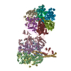
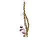

 Mass: 427.201 Da / Num. of mol.: 4 / Source method: obtained synthetically / Formula: C10H15N5O10P2 / Comment: ADP, energy-carrying molecule*YM
 Sample preparation
 / Beamline: BL41XU / Wavelength: 1 Å
 Processing