| Entry | Database: PDB / ID: 4xba
|
|---|
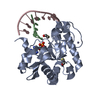
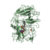
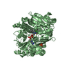
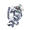
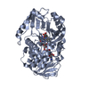
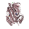
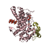
| Title | Hnt3 |
|---|
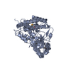
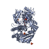
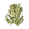
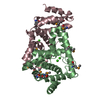
 Components Components | Aprataxin-like protein |
|---|
 Keywords Keywords | HYDROLASE / GMP / nucleotidyl transferase |
|---|
| Function / homology |  Function and homology information Function and homology information
guanosine binding / adenosine-5'-diphospho-5'-[DNA] diphosphatase / DNA-3'-diphospho-5'-guanosine diphosphatase / DNA 5'-adenosine monophosphate hydrolase activity / DNA-3'-diphospho-5'-guanosine diphosphatase activity / single-strand break-containing DNA binding / mismatched DNA binding / single strand break repair / GMP binding / mismatch repair ...guanosine binding / adenosine-5'-diphospho-5'-[DNA] diphosphatase / DNA-3'-diphospho-5'-guanosine diphosphatase / DNA 5'-adenosine monophosphate hydrolase activity / DNA-3'-diphospho-5'-guanosine diphosphatase activity / single-strand break-containing DNA binding / mismatched DNA binding / single strand break repair / GMP binding / mismatch repair / double-stranded RNA binding / single-stranded DNA binding / double-stranded DNA binding / zinc ion binding / nucleus / cytosolSimilarity search - Function Aprataxin, C2HE/C2H2/C2HC zinc finger / C2HE / C2H2 / C2HC zinc-binding finger / HIT domain / HIT-like / HIT-like domain / HIT family, subunit A / HIT-like superfamily / 2-Layer Sandwich / Alpha BetaSimilarity search - Domain/homology |
|---|
| Biological species |   Schizosaccharomyces pombe (fission yeast) Schizosaccharomyces pombe (fission yeast) |
|---|
| Method |  X-RAY DIFFRACTION / SYNCHROTRON / MOLECULAR REPLACEMENT / Resolution: 1.5 Å X-RAY DIFFRACTION / SYNCHROTRON / MOLECULAR REPLACEMENT / Resolution: 1.5 Å |
|---|
 Authors Authors | Jacewicz, A. / Chauleau, M. / Shuman, S. |
|---|
| Funding support |  United States, 1items United States, 1items | Organization | Grant number | Country |
|---|
| National Institutes of Health/National Institute of General Medical Sciences (NIH/NIGMS) | GM46330 | United States |
|
|---|
 Citation Citation | Journal: Nucleic Acids Res. / Year: 2015
Title: DNA3'pp5'G de-capping activity of aprataxin: effect of cap nucleoside analogs and structural basis for guanosine recognition.
Authors: Chauleau, M. / Jacewicz, A. / Shuman, S. |
|---|
| History | | Deposition | Dec 16, 2014 | Deposition site: RCSB / Processing site: RCSB |
|---|
| Revision 1.0 | Jun 3, 2015 | Provider: repository / Type: Initial release |
|---|
| Revision 1.1 | Jun 10, 2015 | Group: Database references |
|---|
| Revision 1.2 | Jul 22, 2015 | Group: Database references |
|---|
| Revision 1.3 | Jul 27, 2016 | Group: Data collection |
|---|
| Revision 1.4 | Sep 13, 2017 | Group: Author supporting evidence / Derived calculations / Category: pdbx_audit_support / pdbx_struct_oper_list
Item: _pdbx_audit_support.funding_organization / _pdbx_struct_oper_list.symmetry_operation |
|---|
| Revision 1.5 | Dec 25, 2019 | Group: Author supporting evidence / Category: pdbx_audit_support / Item: _pdbx_audit_support.funding_organization |
|---|
| Revision 1.6 | Sep 27, 2023 | Group: Data collection / Database references / Refinement description
Category: chem_comp_atom / chem_comp_bond ...chem_comp_atom / chem_comp_bond / database_2 / pdbx_initial_refinement_model
Item: _database_2.pdbx_DOI / _database_2.pdbx_database_accession |
|---|
| Revision 2.0 | Jul 1, 2026 | Group: Atomic model / Data collection / Structure summary
Category: atom_site / chem_comp_atom ...atom_site / chem_comp_atom / chem_comp_bond / pdbx_entry_details
Item: _atom_site.auth_atom_id / _atom_site.label_atom_id ..._atom_site.auth_atom_id / _atom_site.label_atom_id / _chem_comp_atom.atom_id / _chem_comp_bond.atom_id_1 / _chem_comp_bond.atom_id_2 |
|---|
|
|---|
 Movie
Movie Controller


 Open data
 Basic information
 Structure visualization
 Downloads & links
 Other downloads
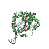
 PDBj
PDBj


 Assembly
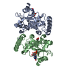

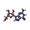


 Mass: 283.241 Da / Num. of mol.: 1 / Source method: obtained synthetically / Formula: C10H13N5O5
 Mass: 65.409 Da / Num. of mol.: 2 / Source method: obtained synthetically / Formula: Zn
 Mass: 92.094 Da / Num. of mol.: 1 / Source method: obtained synthetically / Formula: C3H8O3
 Mass: 363.221 Da / Num. of mol.: 1 / Source method: obtained synthetically / Formula: C10H14N5O8P
 Sample preparation
 Processing