 Movie
Movie Controller

[English] 日本語
 Yorodumi
- PDB-4x1s: The crystal structure of mupain-1-16-D9A in complex with murinise... -
+ Open data

- Basic information
| Entry | Database: PDB / ID: 4x1s | ||||||
|---|---|---|---|---|---|---|---|
| Title | The crystal structure of mupain-1-16-D9A in complex with murinised human uPA at pH7.4 | ||||||
 Components Components |
| ||||||
 Keywords Keywords | HYDROLASE INHIBITOR/HYDROLASE / Serine protease / peptidic inhibitor / uPA / HYDROLASE INHIBITOR-HYDROLASE complex | ||||||
| Function / homology |  Function and homology information Function and homology informationu-plasminogen activator / regulation of smooth muscle cell-matrix adhesion / urokinase plasminogen activator signaling pathway / regulation of plasminogen activation / regulation of integrin-mediated signaling pathway / regulation of fibrinolysis / protein complex involved in cell-matrix adhesion / regulation of wound healing / negative regulation of plasminogen activation / serine-type endopeptidase complex ...u-plasminogen activator / regulation of smooth muscle cell-matrix adhesion / urokinase plasminogen activator signaling pathway / regulation of plasminogen activation / regulation of integrin-mediated signaling pathway / regulation of fibrinolysis / protein complex involved in cell-matrix adhesion / regulation of wound healing / negative regulation of plasminogen activation / serine-type endopeptidase complex / regulation of smooth muscle cell migration / Dissolution of Fibrin Clot / regulation of cell adhesion mediated by integrin / smooth muscle cell migration / plasminogen activation / tertiary granule membrane / negative regulation of fibrinolysis / regulation of cell adhesion / serine protease inhibitor complex / specific granule membrane / fibrinolysis / positive regulation of epidermal growth factor receptor signaling pathway / chemotaxis / blood coagulation / regulation of cell population proliferation / response to hypoxia / positive regulation of cell migration / receptor ligand activity / serine-type endopeptidase activity / external side of plasma membrane / focal adhesion / Neutrophil degranulation / cell surface / signal transduction / proteolysis / : / extracellular exosome / extracellular region / plasma membrane Similarity search - Function | ||||||
| Biological species |  Homo sapiens (human) Homo sapiens (human)synthetic construct (others) | ||||||
| Method |  X-RAY DIFFRACTION / SYNCHROTRON / MOLECULAR REPLACEMENT / Resolution: 1.9 Å X-RAY DIFFRACTION / SYNCHROTRON / MOLECULAR REPLACEMENT / Resolution: 1.9 Å | ||||||
 Authors Authors | Jiang, L. / Zhao, B. / Xu, P. / Andreasen, P. / Huang, M. | ||||||
 Citation Citation | Journal: Plos One / Year: 2014 Title: A cyclic peptidic serine protease inhibitor: increasing affinity by increasing peptide flexibility. Authors: Zhao, B. / Xu, P. / Jiang, L. / Paaske, B. / Kromann-Hansen, T. / Jensen, J.K. / Srensen, H.P. / Liu, Z. / Nielsen, J.T. / Christensen, A. / Hosseini, M. / Srensen, K.K. / Nielsen, N.C. / ...Authors: Zhao, B. / Xu, P. / Jiang, L. / Paaske, B. / Kromann-Hansen, T. / Jensen, J.K. / Srensen, H.P. / Liu, Z. / Nielsen, J.T. / Christensen, A. / Hosseini, M. / Srensen, K.K. / Nielsen, N.C. / Jensen, K.J. / Huang, M. / Andreasen, P.A. | ||||||
| History |
|
- Structure visualization
| Structure viewer | Molecule: MolmilJmol/JSmol |
|---|
- Downloads & links
-Download
| PDBx/mmCIF format | 4x1s.cif.gz | 68.6 KB | Display | PDBx/mmCIF format |
|---|---|---|---|---|
| PDB format | pdb4x1s.ent.gz | 49.5 KB | Display | PDB format |
| PDBx/mmJSON format | 4x1s.json.gz | Tree view | PDBx/mmJSON format | |
| Others |  Other downloads Other downloads |
-Validation report
| Arichive directory | https://data.pdbj.org/pub/pdb/validation_reports/x1/4x1sftp://data.pdbj.org/pub/pdb/validation_reports/x1/4x1s | HTTPS FTP |
|---|
-Related structure data
| Related structure data |  4x1nC  4x1qC  4x1rC  2nwnS C: citing same article ( S: Starting model for refinement |
|---|---|
| Similar structure data |


-Links
 PDBj
PDBj

- Assembly
| Deposited unit | 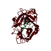
| ||||||||
|---|---|---|---|---|---|---|---|---|---|
| 1 |
| ||||||||
| Unit cell |
|
-Components
| #1: Protein/peptide | Mass: 1061.232 Da / Num. of mol.: 1 / Source method: obtained synthetically / Source: (synth.) synthetic construct (others) |
|---|---|
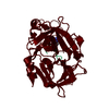
| #2: Protein | Mass: 27869.742 Da / Num. of mol.: 1 / Fragment: catalytic domain (UNP RESIDUES 179-425) / Mutation: H99Y, C122A, N145Q Source method: isolated from a genetically manipulated source Source: (gene. exp.) Homo sapiens (human) / Gene: PLAU / Production host:  Komagataella pastoris (fungus) / References: UniProt: P00749, u-plasminogen activator Komagataella pastoris (fungus) / References: UniProt: P00749, u-plasminogen activator |
| #3: Chemical | ChemComp-MRZ / 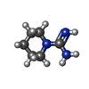  Mass: 127.188 Da / Num. of mol.: 1 / Source method: obtained synthetically / Formula: C6H13N3 Mass: 127.188 Da / Num. of mol.: 1 / Source method: obtained synthetically / Formula: C6H13N3 |
| #4: Water | ChemComp-HOH /  Mass: 18.015 Da / Num. of mol.: 67 / Source method: isolated from a natural source / Formula: H2O Mass: 18.015 Da / Num. of mol.: 67 / Source method: isolated from a natural source / Formula: H2O |
| Has protein modification | Y |
-Experimental details
-Experiment
| Experiment | Method: X-RAY DIFFRACTION |
|---|
- Sample preparation
| Crystal | Density Matthews: 2.11 Å3/Da / Density % sol: 41.63 % |
|---|---|
| Crystal grow | Temperature: 298 K / Method: vapor diffusion, sitting drop / pH: 4.6 Details: 2.0M ammonium sulfate, 50mM sodium citrate pH 4.6, 5% PEG 400 |
-Data collection
| Diffraction | Mean temperature: 100 K |
|---|---|
| Diffraction source | Source: SYNCHROTRON / Site: SSRF  / Beamline: BL17U / Wavelength: 0.979 Å / Beamline: BL17U / Wavelength: 0.979 Å |
| Detector | Type: ADSC QUANTUM 315 / Detector: CCD / Date: Dec 8, 2013 |
| Radiation | Protocol: SINGLE WAVELENGTH / Monochromatic (M) / Laue (L): M / Scattering type: x-ray |
| Radiation wavelength | Wavelength: 0.979 Å / Relative weight: 1 |
| Reflection | Resolution: 1.897→60 Å / Num. obs: 18056 / % possible obs: 97 % / Redundancy: 3.4 % / Net I/σ(I): 32.4 |
- Processing
| Software |
| ||||||||||||||||||||||||||||||||||||||||||||||||||||||||
|---|---|---|---|---|---|---|---|---|---|---|---|---|---|---|---|---|---|---|---|---|---|---|---|---|---|---|---|---|---|---|---|---|---|---|---|---|---|---|---|---|---|---|---|---|---|---|---|---|---|---|---|---|---|---|---|---|---|
| Refinement | Method to determine structure: MOLECULAR REPLACEMENT Starting model: 2NWN Resolution: 1.9→24.11 Å / SU ML: 0.23 / Cross valid method: THROUGHOUT / σ(F): 1.97 / Phase error: 27.97 / Stereochemistry target values: ML
| ||||||||||||||||||||||||||||||||||||||||||||||||||||||||
| Solvent computation | Shrinkage radii: 0.9 Å / VDW probe radii: 1.11 Å / Solvent model: FLAT BULK SOLVENT MODEL | ||||||||||||||||||||||||||||||||||||||||||||||||||||||||
| Refinement step | Cycle: LAST / Resolution: 1.9→24.11 Å
| ||||||||||||||||||||||||||||||||||||||||||||||||||||||||
| Refine LS restraints |
| ||||||||||||||||||||||||||||||||||||||||||||||||||||||||
| LS refinement shell |
|
