[English] 日本語
 Yorodumi
- PDB-6xvd: Crystal structure of complex of urokinase and a upain-1 variant(W... -
+ Open data
- Basic information
| Entry | Database: PDB / ID: 6xvd | |||||||||||||||
|---|---|---|---|---|---|---|---|---|---|---|---|---|---|---|---|---|
| Title | Crystal structure of complex of urokinase and a upain-1 variant(W3F) in pH7.4 condition | |||||||||||||||
 Components Components |
| |||||||||||||||
 Keywords Keywords | PEPTIDE BINDING PROTEIN / upain-1-W3F / urokinase / Cyclic peptide inhibitor | |||||||||||||||
| Function / homology |  Function and homology information Function and homology informationu-plasminogen activator / regulation of smooth muscle cell-matrix adhesion / urokinase plasminogen activator signaling pathway / regulation of plasminogen activation / regulation of fibrinolysis / protein complex involved in cell-matrix adhesion / regulation of signaling receptor activity / regulation of wound healing / negative regulation of plasminogen activation / serine-type endopeptidase complex ...u-plasminogen activator / regulation of smooth muscle cell-matrix adhesion / urokinase plasminogen activator signaling pathway / regulation of plasminogen activation / regulation of fibrinolysis / protein complex involved in cell-matrix adhesion / regulation of signaling receptor activity / regulation of wound healing / negative regulation of plasminogen activation / serine-type endopeptidase complex / regulation of smooth muscle cell migration / Dissolution of Fibrin Clot / smooth muscle cell migration / plasminogen activation / regulation of cell adhesion mediated by integrin / tertiary granule membrane / negative regulation of fibrinolysis / regulation of cell adhesion / specific granule membrane / serine protease inhibitor complex / fibrinolysis / chemotaxis / blood coagulation / regulation of cell population proliferation / response to hypoxia / positive regulation of cell migration / external side of plasma membrane / serine-type endopeptidase activity / focal adhesion / Neutrophil degranulation / cell surface / signal transduction / proteolysis / extracellular space / extracellular exosome / extracellular region / plasma membrane Similarity search - Function | |||||||||||||||
| Biological species |  Homo sapiens (human) Homo sapiens (human)synthetic construct (others) | |||||||||||||||
| Method |  X-RAY DIFFRACTION / X-RAY DIFFRACTION /  SYNCHROTRON / SYNCHROTRON /  MOLECULAR REPLACEMENT / Resolution: 1.4 Å MOLECULAR REPLACEMENT / Resolution: 1.4 Å | |||||||||||||||
 Authors Authors | Xue, G.P. / Xie, X. / Zhou, Y. / Yuan, C. / Huang, M.D. / Jiang, L.G. | |||||||||||||||
| Funding support |  China, 4items China, 4items
| |||||||||||||||
 Citation Citation |  Journal: Biosci.Biotechnol.Biochem. / Year: 2020 Journal: Biosci.Biotechnol.Biochem. / Year: 2020Title: Insight to the residue in P2 position prevents the peptide inhibitor from being hydrolyzed by serine proteases. Authors: Xue, G. / Xie, X. / Zhou, Y. / Yuan, C. / Huang, M. / Jiang, L. | |||||||||||||||
| History |
|
- Structure visualization
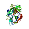
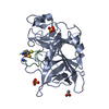
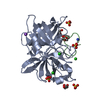
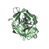
| Structure viewer | Molecule:  Molmil Molmil Jmol/JSmol Jmol/JSmol |
|---|
- Downloads & links
- Download
| PDBx/mmCIF format |  6xvd.cif.gz 6xvd.cif.gz | 69.3 KB | Display |  PDBx/mmCIF format PDBx/mmCIF format |
|---|---|---|---|---|
| PDB format |  pdb6xvd.ent.gz pdb6xvd.ent.gz | 48.8 KB | Display |  PDB format PDB format |
| PDBx/mmJSON format |  6xvd.json.gz 6xvd.json.gz | Tree view |  PDBx/mmJSON format PDBx/mmJSON format | |
| Others |  Other downloads Other downloads |
-Validation report
| Arichive directory |  https://data.pdbj.org/pub/pdb/validation_reports/xv/6xvd https://data.pdbj.org/pub/pdb/validation_reports/xv/6xvd ftp://data.pdbj.org/pub/pdb/validation_reports/xv/6xvd ftp://data.pdbj.org/pub/pdb/validation_reports/xv/6xvd | HTTPS FTP |
|---|
-Related structure data
| Related structure data |  4dvaS S: Starting model for refinement |
|---|---|
| Similar structure data |
- Links
Links
- Assembly
| Deposited unit | 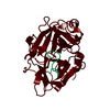
| ||||||||
|---|---|---|---|---|---|---|---|---|---|
| 1 |
| ||||||||
| Unit cell |
|
- Components
| #1: Protein | Mass: 28442.373 Da / Num. of mol.: 1 Source method: isolated from a genetically manipulated source Source: (gene. exp.)  Homo sapiens (human) / Gene: PLAU / Production host: Homo sapiens (human) / Gene: PLAU / Production host:  Komagataella pastoris (fungus) / References: UniProt: P00749, u-plasminogen activator Komagataella pastoris (fungus) / References: UniProt: P00749, u-plasminogen activator |
|---|---|
| #2: Protein/peptide | Mass: 1369.593 Da / Num. of mol.: 1 / Source method: obtained synthetically / Source: (synth.) synthetic construct (others) |
| #3: Water | ChemComp-HOH / |
| Has protein modification | Y |
-Experimental details
-Experiment
| Experiment | Method:  X-RAY DIFFRACTION / Number of used crystals: 1 X-RAY DIFFRACTION / Number of used crystals: 1 |
|---|
- Sample preparation
| Crystal | Density Matthews: 2.04 Å3/Da / Density % sol: 39.76 % |
|---|---|
| Crystal grow | Temperature: 293 K / Method: vapor diffusion, sitting drop / pH: 4.5 Details: 0.05 M sodium citrate at pH 4.5, 1.95 M (NH4)2SO4, 0.05% NaN3, and 5% PEG400 Temp details: room temperature |
-Data collection
| Diffraction | Mean temperature: 100 K / Serial crystal experiment: Y |
|---|---|
| Diffraction source | Source:  SYNCHROTRON / Site: SYNCHROTRON / Site:  SSRF SSRF  / Beamline: BL17U1 / Wavelength: 0.979 Å / Beamline: BL17U1 / Wavelength: 0.979 Å |
| Detector | Type: DECTRIS PILATUS 6M / Detector: PIXEL / Date: Dec 18, 2017 |
| Radiation | Protocol: SINGLE WAVELENGTH / Monochromatic (M) / Laue (L): M / Scattering type: x-ray |
| Radiation wavelength | Wavelength: 0.979 Å / Relative weight: 1 |
| Reflection | Resolution: 1.4→60.65 Å / Num. obs: 46337 / % possible obs: 99.9 % / Redundancy: 5.5 % / Rmerge(I) obs: 0.056 / Net I/σ(I): 25.96 |
| Reflection shell | Resolution: 1.4→1.45 Å / Rmerge(I) obs: 0.056 / Num. unique obs: 46337 |
| Serial crystallography sample delivery | Method: injection |
- Processing
| Software |
| ||||||||||||||||||||||||
|---|---|---|---|---|---|---|---|---|---|---|---|---|---|---|---|---|---|---|---|---|---|---|---|---|---|
| Refinement | Method to determine structure:  MOLECULAR REPLACEMENT MOLECULAR REPLACEMENTStarting model: 4DVA Resolution: 1.4→39.83 Å / Cor.coef. Fo:Fc: 0.958 / Cor.coef. Fo:Fc free: 0.95 / SU B: 0.993 / SU ML: 0.04 / Cross valid method: THROUGHOUT / σ(F): 0 / ESU R: 0.067 / ESU R Free: 0.065 / Stereochemistry target values: MAXIMUM LIKELIHOOD Details: HYDROGENS HAVE BEEN ADDED IN THE RIDING POSITIONS U VALUES : REFINED INDIVIDUALLY
| ||||||||||||||||||||||||
| Solvent computation | Ion probe radii: 0.8 Å / Shrinkage radii: 0.8 Å / VDW probe radii: 1.2 Å / Solvent model: MASK | ||||||||||||||||||||||||
| Displacement parameters | Biso max: 61.06 Å2 / Biso mean: 14.578 Å2 / Biso min: 5.91 Å2
| ||||||||||||||||||||||||
| Refinement step | Cycle: final / Resolution: 1.4→39.83 Å
| ||||||||||||||||||||||||
| LS refinement shell | Resolution: 1.4→1.436 Å / Rfactor Rfree error: 0 / Total num. of bins used: 20
|
 Movie
Movie Controller



 PDBj
PDBj


