| Entry | Database: PDB / ID: 4os6
|
|---|
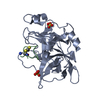
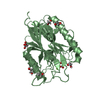
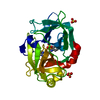
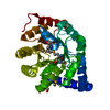
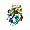
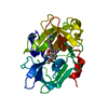
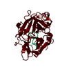
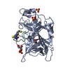
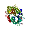
| Title | Crystal structure of urokinase-type plasminogen activator (uPA) complexed with bicyclic peptide UK604 (bicyclic 2) |
|---|
 Components Components | - Urokinase-type plasminogen activator
- bicyclic peptide UK604 (bicyclic 2)
|
|---|
 Keywords Keywords | HYDROLASE/HYDROLASE INHIBITOR / bicyclic peptide / inhibitor / protease / disulfide bridges / cyclization / extracellular / HYDROLASE-HYDROLASE INHIBITOR complex |
|---|
| Function / homology |  Function and homology information Function and homology information
u-plasminogen activator / regulation of smooth muscle cell-matrix adhesion / urokinase plasminogen activator signaling pathway / regulation of plasminogen activation / regulation of integrin-mediated signaling pathway / regulation of fibrinolysis / protein complex involved in cell-matrix adhesion / regulation of wound healing / negative regulation of plasminogen activation / serine-type endopeptidase complex ...u-plasminogen activator / regulation of smooth muscle cell-matrix adhesion / urokinase plasminogen activator signaling pathway / regulation of plasminogen activation / regulation of integrin-mediated signaling pathway / regulation of fibrinolysis / protein complex involved in cell-matrix adhesion / regulation of wound healing / negative regulation of plasminogen activation / serine-type endopeptidase complex / regulation of smooth muscle cell migration / Dissolution of Fibrin Clot / regulation of cell adhesion mediated by integrin / smooth muscle cell migration / plasminogen activation / tertiary granule membrane / negative regulation of fibrinolysis / regulation of cell adhesion / serine protease inhibitor complex / specific granule membrane / fibrinolysis / positive regulation of epidermal growth factor receptor signaling pathway / chemotaxis / blood coagulation / regulation of cell population proliferation / response to hypoxia / positive regulation of cell migration / receptor ligand activity / serine-type endopeptidase activity / external side of plasma membrane / focal adhesion / Neutrophil degranulation / cell surface / signal transduction / proteolysis / : / extracellular exosome / extracellular region / plasma membraneSimilarity search - Function Kringle domain / Kringle / Kringle, conserved site / Kringle superfamily / Kringle domain signature. / Kringle domain profile. / Kringle domain / : / Kringle-like fold / EGF-like domain profile. ...Kringle domain / Kringle / Kringle, conserved site / Kringle superfamily / Kringle domain signature. / Kringle domain profile. / Kringle domain / : / Kringle-like fold / EGF-like domain profile. / EGF-like domain signature 1. / EGF-like domain / Serine proteases, trypsin family, histidine active site / Serine proteases, trypsin family, serine active site / Serine proteases, trypsin family, histidine active site. / Serine proteases, trypsin family, serine active site. / Peptidase S1A, chymotrypsin family / Serine proteases, trypsin domain profile. / Trypsin-like serine protease / Serine proteases, trypsin domain / Trypsin / Trypsin-like serine proteases / Thrombin, subunit H / Peptidase S1, PA clan, chymotrypsin-like fold / Peptidase S1, PA clan / Beta Barrel / Mainly BetaSimilarity search - Domain/homology |
|---|
| Biological species |  Homo sapiens (human) Homo sapiens (human) |
|---|
| Method |  X-RAY DIFFRACTION / SYNCHROTRON / MOLECULAR REPLACEMENT / Resolution: 1.75 Å X-RAY DIFFRACTION / SYNCHROTRON / MOLECULAR REPLACEMENT / Resolution: 1.75 Å |
|---|
 Authors Authors | Chen, S. / Pojer, F. / Heinis, C. |
|---|
 Citation Citation | Journal: Nat Chem / Year: 2014
Title: Dithiol amino acids can structurally shape and enhance the ligand-binding properties of polypeptides.
Authors: Chen, S. / Gopalakrishnan, R. / Schaer, T. / Marger, F. / Hovius, R. / Bertrand, D. / Pojer, F. / Heinis, C. |
|---|
| History | | Deposition | Feb 12, 2014 | Deposition site: RCSB / Processing site: RCSB |
|---|
| Revision 1.0 | Sep 24, 2014 | Provider: repository / Type: Initial release |
|---|
| Revision 1.1 | Nov 5, 2014 | Group: Database references |
|---|
| Revision 1.2 | Nov 22, 2017 | Group: Refinement description / Category: software |
|---|
| Revision 1.3 | Jun 2, 2021 | Group: Database references / Derived calculations / Source and taxonomy
Category: entity_src_gen / struct_conn ...entity_src_gen / struct_conn / struct_ref_seq_dif / struct_site
Item: _entity_src_gen.pdbx_host_org_cell_line / _entity_src_gen.pdbx_host_org_strain ..._entity_src_gen.pdbx_host_org_cell_line / _entity_src_gen.pdbx_host_org_strain / _struct_conn.pdbx_dist_value / _struct_conn.pdbx_leaving_atom_flag / _struct_conn.ptnr1_auth_comp_id / _struct_conn.ptnr1_auth_seq_id / _struct_conn.ptnr1_label_atom_id / _struct_conn.ptnr1_label_comp_id / _struct_conn.ptnr1_label_seq_id / _struct_conn.ptnr2_auth_comp_id / _struct_conn.ptnr2_auth_seq_id / _struct_conn.ptnr2_label_atom_id / _struct_conn.ptnr2_label_comp_id / _struct_conn.ptnr2_label_seq_id / _struct_ref_seq_dif.details / _struct_site.pdbx_auth_asym_id / _struct_site.pdbx_auth_comp_id / _struct_site.pdbx_auth_seq_id |
|---|
| Revision 1.4 | Mar 26, 2025 | Group: Data collection / Database references / Structure summary
Category: chem_comp_atom / chem_comp_bond ...chem_comp_atom / chem_comp_bond / database_2 / pdbx_entry_details / pdbx_modification_feature
Item: _database_2.pdbx_DOI / _database_2.pdbx_database_accession |
|---|
|
|---|
 Movie
Movie Controller

 Yorodumi
 Open data
 Basic information
 Structure visualization
 Downloads & links
 Other downloads


 PDBj
PDBj

 Assembly
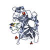

 Type: Cyclic peptide / Class: Inhibitor / Mass: 1478.791 Da / Num. of mol.: 1 / Source method: obtained synthetically / References: bicyclic peptide UK604 (bicyclic 2)
 Mass: 96.063 Da / Num. of mol.: 2 / Source method: obtained synthetically / Formula: SO4
 Mass: 59.044 Da / Num. of mol.: 1 / Source method: obtained synthetically / Formula: C2H3O2
 Mass: 18.015 Da / Num. of mol.: 177 / Source method: isolated from a natural source / Formula: H2O
 Sample preparation
 / Beamline: X06SA / Wavelength: 1 Å
 Processing