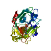
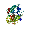
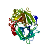
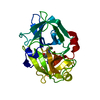
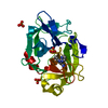
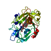
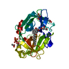
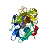
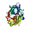
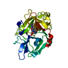
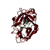
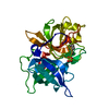
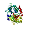
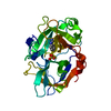
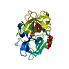
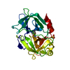
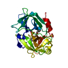
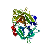
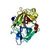
Entry Database : PDB / ID : 4xskTitle Structure of PAItrap, an uPA mutant Urokinase-type plasminogen activator Keywords / / / Function / homology Function Domain/homology Component
/ / / / / / / / / / / / / / / / / / / / / / / / / / / / / / / / / / / / / / / / / / / / / / / / / / / / / / / / / / / / / / / / / / / / / / / / / / / / / / / / / / Biological species Homo sapiens (human)Method / / / Resolution : 1.5 Å Authors Gong, L. / Proulle, V. / Hong, Z. / Lin, Z. / Liu, M. / Yuan, C. / Lin, L. / Furie, B. / Flaumenhaft, R. / Andreasen, P. ...Gong, L. / Proulle, V. / Hong, Z. / Lin, Z. / Liu, M. / Yuan, C. / Lin, L. / Furie, B. / Flaumenhaft, R. / Andreasen, P. / Furie, B. / Huang, M. Funding support Organization Grant number Country National Natural Science Foundation of China 31161130356 NFSC 31370737 National Institutes of Health/National Heart, Lung, and Blood Institute (NIH/NHLBI) HL087203 National Institutes of Health/National Heart, Lung, and Blood Institute (NIH/NHLBI) HL095084 CAS/SFEA International Partnership Program for Creative Research Teams NSF of Fujian Province 2012J05071
Journal : To Be Published Title : Structure of PAItrap, an uPA mutantAuthors : Gong, L. / Proulle, V. / Hong, Z. / Lin, Z. / Liu, M. / Yuan, C. / Lin, L. / Furie, B. / Flaumenhaft, R. / Andreasen, P. / Furie, B. / Huang, M. History Deposition Jan 22, 2015 Deposition site / Processing site Revision 1.0 Feb 3, 2016 Provider / Type Revision 1.1 Oct 18, 2017 Group / Derived calculations / Category / pdbx_struct_oper_listItem / _pdbx_struct_oper_list.symmetry_operationRevision 1.2 Mar 23, 2022 Group / Database references / Category / pdbx_audit_supportItem / _database_2.pdbx_database_accession / _pdbx_audit_support.funding_organizationRevision 1.3 Nov 8, 2023 Group / Refinement descriptionCategory / chem_comp_bond / pdbx_initial_refinement_modelRevision 1.4 Nov 20, 2024 Group / Category / pdbx_modification_feature
Show all Show less
 Movie
Movie Controller


 Open data
 Basic information
 Components
 Keywords
 Function and homology information
 Homo sapiens (human)
 X-RAY DIFFRACTION /
  Authors
 China,
  United States, 6items
  Citation
 Structure visualization
 Downloads & links
 Other downloads
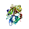

 PDBj
PDBj

 Assembly
 Komagataella pastoris GS115 (fungus) / References: UniProt: P00749, u-plasminogen activator
 Mass: 96.063 Da / Num. of mol.: 2 / Source method: obtained synthetically / Formula: SO4
 Mass: 150.173 Da / Num. of mol.: 2 / Source method: obtained synthetically / Formula: C6H14O4
 Mass: 92.094 Da / Num. of mol.: 2 / Source method: obtained synthetically / Formula: C3H8O3
 Mass: 18.015 Da / Num. of mol.: 121 / Source method: isolated from a natural source / Formula: H2O
 Sample preparation
 Processing