 Movie
Movie Controller


+ Open data

- Basic information
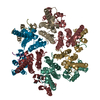
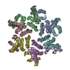
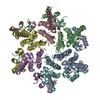
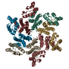
| Entry | Database: PDB / ID: 4wym | ||||||
|---|---|---|---|---|---|---|---|
| Title | Structural basis of HIV-1 capsid recognition by CPSF6 | ||||||
 Components Components |
| ||||||
 Keywords Keywords | VIRAL PROTEIN / CAPSID PROTEIN / HEXAMER / ENGINEERED DISULFIDE BONDS / CPSF6 / viral restriction | ||||||
| Function / homology |  Function and homology information Function and homology informationexon-exon junction complex binding / mRNA alternative polyadenylation / positive regulation of RNA export from nucleus / mRNA cleavage factor complex / co-transcriptional mRNA 3'-end processing, cleavage and polyadenylation pathway / interchromatin granule / perichromatin fibrils / Processing of Intronless Pre-mRNAs / mRNA cleavage and polyadenylation specificity factor complex / mRNA 3'-end processing ...exon-exon junction complex binding / mRNA alternative polyadenylation / positive regulation of RNA export from nucleus / mRNA cleavage factor complex / co-transcriptional mRNA 3'-end processing, cleavage and polyadenylation pathway / interchromatin granule / perichromatin fibrils / Processing of Intronless Pre-mRNAs / mRNA cleavage and polyadenylation specificity factor complex / mRNA 3'-end processing / Signaling by cytosolic FGFR1 fusion mutants / paraspeckles / mRNA 3'-end processing / RNA Polymerase II Transcription Termination / protein heterotetramerization / viral budding via host ESCRT complex / ribosomal large subunit binding / Processing of Capped Intron-Containing Pre-mRNA / Signaling by FGFR1 in disease / HIV-1 retropepsin / symbiont-mediated activation of host apoptosis / retroviral ribonuclease H / exoribonuclease H / exoribonuclease H activity / protein tetramerization / DNA integration / viral genome integration into host DNA / establishment of integrated proviral latency / RNA-directed DNA polymerase / RNA stem-loop binding / viral penetration into host nucleus / host multivesicular body / RNA-directed DNA polymerase activity / RNA-DNA hybrid ribonuclease activity / ISG15 antiviral mechanism / Transferases; Transferring phosphorus-containing groups; Nucleotidyltransferases / mRNA processing / host cell / viral nucleocapsid / DNA recombination / DNA-directed DNA polymerase / aspartic-type endopeptidase activity / Hydrolases; Acting on ester bonds / DNA-directed DNA polymerase activity / nuclear speck / ribonucleoprotein complex / symbiont-mediated suppression of host gene expression / viral translational frameshifting / mRNA binding / symbiont entry into host cell / lipid binding / host cell nucleus / host cell plasma membrane / virion membrane / structural molecule activity / proteolysis / DNA binding / RNA binding / zinc ion binding / nucleoplasm / membrane / nucleus / cytoplasm Similarity search - Function | ||||||
| Biological species |  Human immunodeficiency virus type 1 group M subtype B Human immunodeficiency virus type 1 group M subtype B Homo sapiens (human) Homo sapiens (human) | ||||||
| Method |  X-RAY DIFFRACTION / SYNCHROTRON / FOURIER SYNTHESIS / Resolution: 2.6 Å X-RAY DIFFRACTION / SYNCHROTRON / FOURIER SYNTHESIS / Resolution: 2.6 Å | ||||||
 Authors Authors | Battacharya, A. / Taylor, A.B. / Hart, P.J. / Ivanov, D.N. | ||||||
 Citation Citation | Journal: Proc.Natl.Acad.Sci.USA / Year: 2014 Title: Structural basis of HIV-1 capsid recognition by PF74 and CPSF6. Authors: Bhattacharya, A. / Alam, S.L. / Fricke, T. / Zadrozny, K. / Sedzicki, J. / Taylor, A.B. / Demeler, B. / Pornillos, O. / Ganser-Pornillos, B.K. / Diaz-Griffero, F. / Ivanov, D.N. / Yeager, M. | ||||||
| History |
|
- Structure visualization
| Structure viewer | Molecule: MolmilJmol/JSmol |
|---|
- Downloads & links
-Download
| PDBx/mmCIF format | 4wym.cif.gz | 506.7 KB | Display | PDBx/mmCIF format |
|---|---|---|---|---|
| PDB format | pdb4wym.ent.gz | 422.8 KB | Display | PDB format |
| PDBx/mmJSON format | 4wym.json.gz | Tree view | PDBx/mmJSON format | |
| Others |  Other downloads Other downloads |
-Validation report
| Arichive directory | https://data.pdbj.org/pub/pdb/validation_reports/wy/4wymftp://data.pdbj.org/pub/pdb/validation_reports/wy/4wym | HTTPS FTP |
|---|
-Related structure data
| Related structure data |  4qnbC 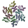 3h4eS C: citing same article ( S: Starting model for refinement |
|---|---|
| Similar structure data |


-Links
 PDBj
PDBj


- Assembly
| Deposited unit | 
| ||||||||
|---|---|---|---|---|---|---|---|---|---|
| 1 | 
| ||||||||
| 2 | 
| ||||||||
| Unit cell |
|
-Components
| #1: Protein | Mass: 25461.271 Da / Num. of mol.: 12 / Mutation: A14C, E45C, W184A, M185A Source method: isolated from a genetically manipulated source Source: (gene. exp.) Human immunodeficiency virus type 1 group M subtype BStrain: isolate NY5 / Gene: gag / Plasmid: PET11A / Production host:  #2: Protein/peptide | Mass: 1708.952 Da / Num. of mol.: 11 / Fragment: UNP RESIDUES 313-327 Source method: isolated from a genetically manipulated source Source: (gene. exp.) Homo sapiens (human) / Gene: CPSF6, CFIM68 / Plasmid: pET30a / Production host: Has protein modification | Y | |
|---|
-Experimental details
-Experiment
| Experiment | Method: X-RAY DIFFRACTION |
|---|
- Sample preparation
| Crystal | Density Matthews: 2.96 Å3/Da / Density % sol: 58.42 % |
|---|---|
| Crystal grow | Temperature: 295 K / Method: vapor diffusion, sitting drop / pH: 7.5 Details: 0.1 M sodium formate, ammonium acetate, tri-sodium citrate, sodium/potassium tartrate, sodium oxamate; 0.1 M sodium HEPES, MOPS; 30% glycerol, PEG 4000 |
-Data collection
| Diffraction | Mean temperature: 100 K |
|---|---|
| Diffraction source | Source: SYNCHROTRON / Site: APS  / Beamline: 24-ID-C / Wavelength: 0.9795 Å / Beamline: 24-ID-C / Wavelength: 0.9795 Å |
| Detector | Type: PSI PILATUS 6M / Detector: PIXEL / Date: Jul 2, 2013 |
| Radiation | Protocol: SINGLE WAVELENGTH / Monochromatic (M) / Laue (L): M / Scattering type: x-ray |
| Radiation wavelength | Wavelength: 0.9795 Å / Relative weight: 1 |
| Reflection | Resolution: 2.6→48.47 Å / Num. obs: 117525 / % possible obs: 100 % / Redundancy: 6.8 % / Biso Wilson estimate: 62.65 Å2 / Rsym value: 0.065 / Net I/σ(I): 20.2 |
| Reflection shell | Resolution: 2.6→2.74 Å / Redundancy: 7.1 % / Rmerge(I) obs: 0.863 / Mean I/σ(I) obs: 2.3 / % possible all: 100 |
- Processing
| Software |
| |||||||||||||||||||||||||||||||||||||||||||||||||||||||||||||||||||||||||||||||||||||||||||||||||||||||||
|---|---|---|---|---|---|---|---|---|---|---|---|---|---|---|---|---|---|---|---|---|---|---|---|---|---|---|---|---|---|---|---|---|---|---|---|---|---|---|---|---|---|---|---|---|---|---|---|---|---|---|---|---|---|---|---|---|---|---|---|---|---|---|---|---|---|---|---|---|---|---|---|---|---|---|---|---|---|---|---|---|---|---|---|---|---|---|---|---|---|---|---|---|---|---|---|---|---|---|---|---|---|---|---|---|---|---|
| Refinement | Method to determine structure: FOURIER SYNTHESIS Starting model: 3H4E Resolution: 2.6→48.468 Å / SU ML: 0.36 / Cross valid method: THROUGHOUT / σ(F): 1.91 / Phase error: 28.16 / Stereochemistry target values: ML
| |||||||||||||||||||||||||||||||||||||||||||||||||||||||||||||||||||||||||||||||||||||||||||||||||||||||||
| Solvent computation | Shrinkage radii: 0.9 Å / VDW probe radii: 1.11 Å / Solvent model: FLAT BULK SOLVENT MODEL | |||||||||||||||||||||||||||||||||||||||||||||||||||||||||||||||||||||||||||||||||||||||||||||||||||||||||
| Displacement parameters | Biso mean: 73.97 Å2 | |||||||||||||||||||||||||||||||||||||||||||||||||||||||||||||||||||||||||||||||||||||||||||||||||||||||||
| Refinement step | Cycle: LAST / Resolution: 2.6→48.468 Å
| |||||||||||||||||||||||||||||||||||||||||||||||||||||||||||||||||||||||||||||||||||||||||||||||||||||||||
| Refine LS restraints |
| |||||||||||||||||||||||||||||||||||||||||||||||||||||||||||||||||||||||||||||||||||||||||||||||||||||||||
| LS refinement shell |
|
