 Movie
Movie Controller

[English] 日本語
 Yorodumi
- PDB-4qnb: Disulfide stabilized HIV-1 CA hexamer in complex with PHENYL-L-PH... -
+ Open data

- Basic information
| Entry | Database: PDB / ID: 4qnb | ||||||
|---|---|---|---|---|---|---|---|
| Title | Disulfide stabilized HIV-1 CA hexamer in complex with PHENYL-L-PHENYLALANINAMIDE inhibitor | ||||||
 Components Components | CAPSID PROTEIN P24 | ||||||
 Keywords Keywords | VIRAL PROTEIN/INHIBITOR / capsid protein / disulfide crosslink / VIRAL PROTEIN / VIRAL PROTEIN-INHIBITOR complex | ||||||
| Function / homology |  Function and homology information Function and homology informationHIV-1 retropepsin / symbiont-mediated activation of host apoptosis / retroviral ribonuclease H / exoribonuclease H / exoribonuclease H activity / DNA integration / viral genome integration into host DNA / establishment of integrated proviral latency / RNA-directed DNA polymerase / RNA stem-loop binding ...HIV-1 retropepsin / symbiont-mediated activation of host apoptosis / retroviral ribonuclease H / exoribonuclease H / exoribonuclease H activity / DNA integration / viral genome integration into host DNA / establishment of integrated proviral latency / RNA-directed DNA polymerase / RNA stem-loop binding / viral penetration into host nucleus / host multivesicular body / RNA-directed DNA polymerase activity / RNA-DNA hybrid ribonuclease activity / Transferases; Transferring phosphorus-containing groups; Nucleotidyltransferases / host cell / viral nucleocapsid / DNA recombination / DNA-directed DNA polymerase / aspartic-type endopeptidase activity / Hydrolases; Acting on ester bonds / DNA-directed DNA polymerase activity / symbiont-mediated suppression of host gene expression / viral translational frameshifting / symbiont entry into host cell / lipid binding / host cell nucleus / host cell plasma membrane / virion membrane / structural molecule activity / proteolysis / DNA binding / zinc ion binding Similarity search - Function | ||||||
| Biological species |   Human immunodeficiency virus type 1 Human immunodeficiency virus type 1 | ||||||
| Method |  X-RAY DIFFRACTION / SYNCHROTRON / MOLECULAR REPLACEMENT / Resolution: 1.996 Å X-RAY DIFFRACTION / SYNCHROTRON / MOLECULAR REPLACEMENT / Resolution: 1.996 Å | ||||||
 Authors Authors | Pornillos, O. | ||||||
 Citation Citation | Journal: Proc.Natl.Acad.Sci.USA / Year: 2014 Title: Structural basis of HIV-1 capsid recognition by PF74 and CPSF6. Authors: Bhattacharya, A. / Alam, S.L. / Fricke, T. / Zadrozny, K. / Sedzicki, J. / Taylor, A.B. / Demeler, B. / Pornillos, O. / Ganser-Pornillos, B.K. / Diaz-Griffero, F. / Ivanov, D.N. / Yeager, M. | ||||||
| History |
|
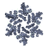
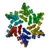
- Structure visualization
| Structure viewer | Molecule: MolmilJmol/JSmol |
|---|
- Downloads & links
-Download
| PDBx/mmCIF format | 4qnb.cif.gz | 102.7 KB | Display | PDBx/mmCIF format |
|---|---|---|---|---|
| PDB format | pdb4qnb.ent.gz | 78.6 KB | Display | PDB format |
| PDBx/mmJSON format | 4qnb.json.gz | Tree view | PDBx/mmJSON format | |
| Others |  Other downloads Other downloads |
-Validation report
| Arichive directory | https://data.pdbj.org/pub/pdb/validation_reports/qn/4qnbftp://data.pdbj.org/pub/pdb/validation_reports/qn/4qnb | HTTPS FTP |
|---|
-Related structure data
| Related structure data |  4wymC  3h47S S: Starting model for refinement C: citing same article ( |
|---|---|
| Similar structure data |


-Links
 PDBj
PDBj
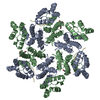
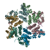
- Assembly
| Deposited unit | 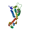
| ||||||||
|---|---|---|---|---|---|---|---|---|---|
| 1 | x 6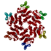
| ||||||||
| Unit cell |
|
-Components
| #1: Protein | Mass: 25461.271 Da / Num. of mol.: 1 / Mutation: A14C,E45C,W184A,M185A Source method: isolated from a genetically manipulated source Source: (gene. exp.) Human immunodeficiency virus type 1 (NEW YORK-5 ISOLATE)Gene: gag-pol / Plasmid: pET11a / Production host:  |
|---|---|
| #2: Chemical | ChemComp-1B0 / 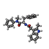  Mass: 425.522 Da / Num. of mol.: 1 / Source method: obtained synthetically / Formula: C27H27N3O2 Mass: 425.522 Da / Num. of mol.: 1 / Source method: obtained synthetically / Formula: C27H27N3O2 |
| #3: Water | ChemComp-HOH /  Mass: 18.015 Da / Num. of mol.: 100 / Source method: isolated from a natural source / Formula: H2O Mass: 18.015 Da / Num. of mol.: 100 / Source method: isolated from a natural source / Formula: H2O |
| Has protein modification | Y |
-Experimental details
-Experiment
| Experiment | Method: X-RAY DIFFRACTION |
|---|
- Sample preparation
| Crystal | Density Matthews: 2.68 Å3/Da / Density % sol: 54.08 % |
|---|---|
| Crystal grow | Temperature: 293 K / Method: vapor diffusion, sitting drop / pH: 8 Details: 10% PEG 8,000, 2% Tacsimate, 100 mM Tris, VAPOR DIFFUSION, SITTING DROP, temperature 293K |
-Data collection
| Diffraction | Mean temperature: 100 K |
|---|---|
| Diffraction source | Source: SYNCHROTRON / Site: APS  / Beamline: 22-BM / Wavelength: 1 Å / Beamline: 22-BM / Wavelength: 1 Å |
| Detector | Type: MAR scanner 345 mm plate / Detector: IMAGE PLATE / Date: Jan 11, 2011 |
| Radiation | Protocol: SINGLE WAVELENGTH / Monochromatic (M) / Laue (L): M / Scattering type: x-ray |
| Radiation wavelength | Wavelength: 1 Å / Relative weight: 1 |
| Reflection | Resolution: 1.996→50 Å / Num. obs: 18424 / Observed criterion σ(F): 2 / Observed criterion σ(I): 2 / Redundancy: 11.1 % / Biso Wilson estimate: 28 Å2 / Rsym value: 0.087 / Net I/σ(I): 43.9 |
- Processing
| Software |
| ||||||||||||||||||||||||||||||||||||||||||
|---|---|---|---|---|---|---|---|---|---|---|---|---|---|---|---|---|---|---|---|---|---|---|---|---|---|---|---|---|---|---|---|---|---|---|---|---|---|---|---|---|---|---|---|
| Refinement | Method to determine structure: MOLECULAR REPLACEMENT Starting model: 3h47 Resolution: 1.996→26.642 Å / SU ML: 0.22 / σ(F): 1.34 / Phase error: 26.53 / Stereochemistry target values: ML
| ||||||||||||||||||||||||||||||||||||||||||
| Solvent computation | Shrinkage radii: 0.9 Å / VDW probe radii: 1.11 Å / Solvent model: FLAT BULK SOLVENT MODEL | ||||||||||||||||||||||||||||||||||||||||||
| Refinement step | Cycle: LAST / Resolution: 1.996→26.642 Å
| ||||||||||||||||||||||||||||||||||||||||||
| Refine LS restraints |
| ||||||||||||||||||||||||||||||||||||||||||
| LS refinement shell | Refine-ID: X-RAY DIFFRACTION / % reflection obs: 100 %
| ||||||||||||||||||||||||||||||||||||||||||
| Refinement TLS params. | Method: refined / Origin x: 24.8716 Å / Origin y: 10.3507 Å / Origin z: -0.7647 Å
| ||||||||||||||||||||||||||||||||||||||||||
| Refinement TLS group | Selection details: all |
