Monochromator: SI(111) / Protocol: SINGLE WAVELENGTH / Monochromatic (M) / Laue (L): M / Scattering type: x-ray
Radiation wavelength
Wavelength: 0.9793 Å / Relative weight: 1
Reflection
Resolution: 1.09→25 Å / Num. obs: 88243 / % possible obs: 99.6 % / Observed criterion σ(I): -3 / Redundancy: 9.6 % / Rmerge(I) obs: 0.066 / Net I/σ(I): 32.6
Reflection shell
Resolution: 1.09→1.11 Å / Redundancy: 6.4 % / Rmerge(I) obs: 0.795 / Mean I/σ(I) obs: 2.4 / % possible all: 95.7
-
Processing
Software
Name
Version
Classification
PHENIX
(phenix.refine: 1.9_1692)
refinement
HKL-3000
phasing
HKL-3000
datascaling
SBC-Collect
datacollection
SHARP
phasing
ARP
modelbuilding
Coot
modelbuilding
Refinement
Method to determine structure: MAD / Resolution: 1.09→24.712 Å / SU ML: 0.08 / Cross valid method: THROUGHOUT / σ(F): 0 / Phase error: 11.89 / Stereochemistry target values: ML Details: Hydrogen atoms were added in riding positions. The mFo-DFc difference map shows a strong peak near His82, which is possibly a disordered citrate molecule.
Rfactor
Num. reflection
% reflection
Rfree
0.1376
1782
2.02 %
Rwork
0.1279
-
-
obs
0.1281
88155
99.35 %
Solvent computation
Shrinkage radii: 0.9 Å / VDW probe radii: 1.11 Å / Solvent model: FLAT BULK SOLVENT MODEL
Refinement step
Cycle: LAST / Resolution: 1.09→24.712 Å
Protein
Nucleic acid
Ligand
Solvent
Total
Num. atoms
1496
0
39
260
1795
Refine LS restraints
Refine-ID
Type
Dev ideal
Number
X-RAY DIFFRACTION
f_bond_d
0.011
1676
X-RAY DIFFRACTION
f_angle_d
1.514
2326
X-RAY DIFFRACTION
f_dihedral_angle_d
16.783
586
X-RAY DIFFRACTION
f_chiral_restr
0.092
269
X-RAY DIFFRACTION
f_plane_restr
0.01
314
LS refinement shell
Resolution (Å)
Rfactor Rfree
Num. reflection Rfree
Rfactor Rwork
Num. reflection Rwork
Refine-ID
% reflection obs (%)
1.09-1.1399
0.2196
218
0.2118
10215
X-RAY DIFFRACTION
95
1.1399-1.2
0.1505
217
0.1453
10708
X-RAY DIFFRACTION
100
1.2-1.2752
0.1353
224
0.1223
10769
X-RAY DIFFRACTION
100
1.2752-1.3736
0.1361
222
0.1118
10787
X-RAY DIFFRACTION
100
1.3736-1.5119
0.1084
218
0.0962
10819
X-RAY DIFFRACTION
100
1.5119-1.7306
0.1208
233
0.0942
10847
X-RAY DIFFRACTION
100
1.7306-2.1802
0.1273
219
0.1113
10957
X-RAY DIFFRACTION
100
2.1802-24.7178
0.1446
231
0.1446
11271
X-RAY DIFFRACTION
100
+
About Yorodumi
-
News
-
Feb 9, 2022. New format data for meta-information of EMDB entries
New format data for meta-information of EMDB entries
Version 3 of the EMDB header file is now the official format.
The previous official version 1.9 will be removed from the archive.
In the structure databanks used in Yorodumi, some data are registered as the other names, "COVID-19 virus" and "2019-nCoV". Here are the details of the virus and the list of structure data.
Jan 31, 2019. EMDB accession codes are about to change! (news from PDBe EMDB page)
EMDB accession codes are about to change! (news from PDBe EMDB page)
The allocation of 4 digits for EMDB accession codes will soon come to an end. Whilst these codes will remain in use, new EMDB accession codes will include an additional digit and will expand incrementally as the available range of codes is exhausted. The current 4-digit format prefixed with “EMD-” (i.e. EMD-XXXX) will advance to a 5-digit format (i.e. EMD-XXXXX), and so on. It is currently estimated that the 4-digit codes will be depleted around Spring 2019, at which point the 5-digit format will come into force.
The EM Navigator/Yorodumi systems omit the EMD- prefix.
Related info.:Q: What is EMD? / ID/Accession-code notation in Yorodumi/EM Navigator
Yorodumi is a browser for structure data from EMDB, PDB, SASBDB, etc.
This page is also the successor to EM Navigator detail page, and also detail information page/front-end page for Omokage search.
The word "yorodu" (or yorozu) is an old Japanese word meaning "ten thousand". "mi" (miru) is to see.
Related info.:EMDB / PDB / SASBDB / Comparison of 3 databanks / Yorodumi Search / Aug 31, 2016. New EM Navigator & Yorodumi / Yorodumi Papers / Jmol/JSmol / Function and homology information / Changes in new EM Navigator and Yorodumi
 Movie
Movie Controller


 Open data
 Basic information
 Components
 Keywords
 Function and homology information
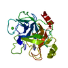
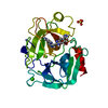
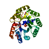
 Jonesia denitrificans (bacteria)
 X-RAY DIFFRACTION /
  Authors
 United States, 1items
  Citation
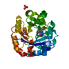
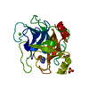
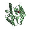
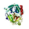
 Structure visualization
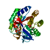
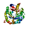
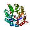
 Downloads & links
 Other downloads
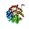


 PDBj
PDBj
 Assembly

 Mass: 192.124 Da / Num. of mol.: 3 / Source method: obtained synthetically / Formula: C6H8O7
 Mass: 18.015 Da / Num. of mol.: 260 / Source method: isolated from a natural source / Formula: H2O
 Sample preparation
 Processing