 Movie
Movie Controller

[English] 日本語
 Yorodumi
- PDB-5xsf: Crystal structure of the 2-keto-3-deoxy-6-phosphogluconate aldola... -
+ Open data

- Basic information
| Entry | Database: PDB / ID: 5xsf | ||||||
|---|---|---|---|---|---|---|---|
| Title | Crystal structure of the 2-keto-3-deoxy-6-phosphogluconate aldolase of Zymomonas mobilis ZM4 with 3-phosphoglycerate | ||||||
 Components Components | KHG/KDPG aldolase | ||||||
 Keywords Keywords | LYASE | ||||||
| Function / homology |  Function and homology information Function and homology information2-dehydro-3-deoxy-phosphogluconate aldolase / 2-dehydro-3-deoxy-phosphogluconate aldolase activity / cytoplasm Similarity search - Function | ||||||
| Biological species |  Zymomonas mobilis subsp. mobilis (bacteria) Zymomonas mobilis subsp. mobilis (bacteria) | ||||||
| Method |  X-RAY DIFFRACTION / SYNCHROTRON / MOLECULAR REPLACEMENT / Resolution: 1.962 Å X-RAY DIFFRACTION / SYNCHROTRON / MOLECULAR REPLACEMENT / Resolution: 1.962 Å | ||||||
 Authors Authors | Seo, P.W. / Kim, J.S. | ||||||
 Citation Citation | Journal: To Be Published Title: Crystal structure of the 2-keto-3-deoxy-6-phosphogluconate aldolase of Zymomonas mobilis ZM4 Authors: Seo, P.W. / Kim, J.S. | ||||||
| History |
|
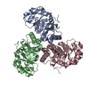
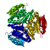
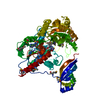
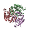
- Structure visualization
| Structure viewer | Molecule: MolmilJmol/JSmol |
|---|
- Downloads & links
-Download
| PDBx/mmCIF format | 5xsf.cif.gz | 99.2 KB | Display | PDBx/mmCIF format |
|---|---|---|---|---|
| PDB format | pdb5xsf.ent.gz | 77.1 KB | Display | PDB format |
| PDBx/mmJSON format | 5xsf.json.gz | Tree view | PDBx/mmJSON format | |
| Others |  Other downloads Other downloads |
-Validation report
| Arichive directory | https://data.pdbj.org/pub/pdb/validation_reports/xs/5xsfftp://data.pdbj.org/pub/pdb/validation_reports/xs/5xsf | HTTPS FTP |
|---|
-Related structure data
| Related structure data |  5xseSC S: Starting model for refinement C: citing same article ( |
|---|---|
| Similar structure data |
-Links
 PDBj
PDBj
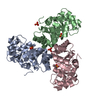
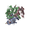
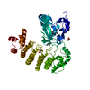
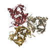
- Assembly
| Deposited unit | 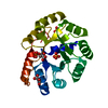
| ||||||||||||||||||
|---|---|---|---|---|---|---|---|---|---|---|---|---|---|---|---|---|---|---|---|
| 1 | 
| ||||||||||||||||||
| Unit cell |
| ||||||||||||||||||
| Components on special symmetry positions |
|
-Components
| #1: Protein | Mass: 21597.402 Da / Num. of mol.: 1 Source method: isolated from a genetically manipulated source Source: (gene. exp.) Zymomonas mobilis subsp. mobilis (strain ATCC 31821 / ZM4 / CP4) (bacteria)Strain: ATCC 31821 / ZM4 / CP4 / Gene: eda, kdgA, ZMO0997 / Production host: References: UniProt: Q00384, 2-dehydro-3-deoxy-phosphogluconate aldolase | ||||||
|---|---|---|---|---|---|---|---|
| #2: Chemical |   Mass: 94.971 Da / Num. of mol.: 2 / Source method: obtained synthetically / Formula: PO4 / Feature type: SUBJECT OF INVESTIGATION Mass: 94.971 Da / Num. of mol.: 2 / Source method: obtained synthetically / Formula: PO4 / Feature type: SUBJECT OF INVESTIGATION#3: Chemical | ChemComp-3PG / |   Mass: 186.057 Da / Num. of mol.: 1 / Source method: obtained synthetically / Formula: C3H7O7P / Feature type: SUBJECT OF INVESTIGATION Mass: 186.057 Da / Num. of mol.: 1 / Source method: obtained synthetically / Formula: C3H7O7P / Feature type: SUBJECT OF INVESTIGATION#4: Chemical | ChemComp-3PY / |   Mass: 104.061 Da / Num. of mol.: 1 / Source method: obtained synthetically / Formula: C3H4O4 / Feature type: SUBJECT OF INVESTIGATION Mass: 104.061 Da / Num. of mol.: 1 / Source method: obtained synthetically / Formula: C3H4O4 / Feature type: SUBJECT OF INVESTIGATION#5: Water | ChemComp-HOH / |  Mass: 18.015 Da / Num. of mol.: 150 / Source method: isolated from a natural source / Formula: H2O Mass: 18.015 Da / Num. of mol.: 150 / Source method: isolated from a natural source / Formula: H2O |
-Experimental details
-Experiment
| Experiment | Method: X-RAY DIFFRACTION / Number of used crystals: 1 |
|---|
- Sample preparation
| Crystal | Density Matthews: 3.12 Å3/Da / Density % sol: 60.56 % |
|---|---|
| Crystal grow | Temperature: 291 K / Method: vapor diffusion, hanging drop Details: 1.6M Ammonium Sulfate, 0.1M CAPS (pH 10.5), 0.2M Lithium Sulfate |
-Data collection
| Diffraction | Mean temperature: 100 K |
|---|---|
| Diffraction source | Source: SYNCHROTRON / Site: PAL/PLS  / Beamline: 7A (6B, 6C1) / Wavelength: 1 Å / Beamline: 7A (6B, 6C1) / Wavelength: 1 Å |
| Detector | Type: ADSC QUANTUM 270 / Detector: CCD / Date: Jun 14, 2016 |
| Radiation | Protocol: SINGLE WAVELENGTH / Monochromatic (M) / Laue (L): M / Scattering type: x-ray |
| Radiation wavelength | Wavelength: 1 Å / Relative weight: 1 |
| Reflection | Resolution: 1.962→35.853 Å / Num. obs: 20372 / % possible obs: 99.5 % / Redundancy: 13.2 % / Net I/σ(I): 17.8 |
| Reflection shell | Resolution: 1.96→1.99 Å / Redundancy: 18.3 % / Mean I/σ(I) obs: 3.4 / Rsym value: 0.399 / % possible all: 100 |
- Processing
| Software |
| |||||||||||||||||||||||||||||||||||||||||||||||||
|---|---|---|---|---|---|---|---|---|---|---|---|---|---|---|---|---|---|---|---|---|---|---|---|---|---|---|---|---|---|---|---|---|---|---|---|---|---|---|---|---|---|---|---|---|---|---|---|---|---|---|
| Refinement | Method to determine structure: MOLECULAR REPLACEMENT Starting model: 5xse Resolution: 1.962→35.853 Å / SU ML: 0.16 / Cross valid method: FREE R-VALUE / σ(F): 1.54 / Phase error: 18.58
| |||||||||||||||||||||||||||||||||||||||||||||||||
| Solvent computation | Shrinkage radii: 0.9 Å / VDW probe radii: 1.11 Å | |||||||||||||||||||||||||||||||||||||||||||||||||
| Refinement step | Cycle: LAST / Resolution: 1.962→35.853 Å
| |||||||||||||||||||||||||||||||||||||||||||||||||
| Refine LS restraints |
| |||||||||||||||||||||||||||||||||||||||||||||||||
| LS refinement shell |
| |||||||||||||||||||||||||||||||||||||||||||||||||
| Refinement TLS params. | Method: refined / Origin x: 6.9983 Å / Origin y: 25.1353 Å / Origin z: -34.2592 Å
| |||||||||||||||||||||||||||||||||||||||||||||||||
| Refinement TLS group | Selection details: all |
