Resolution: 2.15→2.23 Å / Redundancy: 6.5 % / Rmerge(I) obs: 0.492 / Mean I/σ(I) obs: 4.9 / Num. unique all: 5796 / % possible all: 99.8
-
Processing
Software
Name
Version
Classification
ADSC
Quantum
datacollection
SHELXDE
phasing
PHENIX
(phenix.refine: 1.6_289)
refinement
DENZO
datareduction
SCALEPACK
datascaling
Refinement
Method to determine structure: SAD / Resolution: 2.15→36.49 Å / SU ML: 0.4 / Cross valid method: THROUGHOUT / σ(F): 0.92 / Phase error: 30.45 / Stereochemistry target values: ML
Rfactor
Num. reflection
% reflection
Selection details
Rfree
0.2709
2868
5.04 %
RANDOM
Rwork
0.2153
-
-
-
obs
0.2182
56907
98.19 %
-
Solvent computation
Shrinkage radii: 0.9 Å / VDW probe radii: 1.11 Å / Solvent model: FLAT BULK SOLVENT MODEL / Bsol: 41.271 Å2 / ksol: 0.338 e/Å3
Displacement parameters
Baniso -1
Baniso -2
Baniso -3
1-
-9.4844 Å2
0 Å2
-0 Å2
2-
-
13.4429 Å2
-0 Å2
3-
-
-
-3.9585 Å2
Refinement step
Cycle: LAST / Resolution: 2.15→36.49 Å
Protein
Nucleic acid
Ligand
Solvent
Total
Num. atoms
4060
0
53
112
4225
Refine LS restraints
Refine-ID
Type
Dev ideal
Number
X-RAY DIFFRACTION
f_bond_d
0.009
4202
X-RAY DIFFRACTION
f_angle_d
1.21
5691
X-RAY DIFFRACTION
f_dihedral_angle_d
18.916
1564
X-RAY DIFFRACTION
f_chiral_restr
0.08
622
X-RAY DIFFRACTION
f_plane_restr
0.006
745
LS refinement shell
Resolution (Å)
Rfactor Rfree
Num. reflection Rfree
Rfactor Rwork
Num. reflection Rwork
Refine-ID
% reflection obs (%)
2.1502-2.1872
0.3672
111
0.3091
2671
X-RAY DIFFRACTION
97
2.1872-2.227
0.3757
149
0.2677
2769
X-RAY DIFFRACTION
100
2.227-2.2698
0.3395
171
0.2516
2737
X-RAY DIFFRACTION
100
2.2698-2.3162
0.2702
130
0.2493
2772
X-RAY DIFFRACTION
100
2.3162-2.3665
0.3148
151
0.2328
2754
X-RAY DIFFRACTION
100
2.3665-2.4215
0.3081
164
0.2369
2701
X-RAY DIFFRACTION
100
2.4215-2.4821
0.3432
157
0.2532
2752
X-RAY DIFFRACTION
100
2.4821-2.5492
0.3512
138
0.2673
2756
X-RAY DIFFRACTION
100
2.5492-2.6242
0.3632
141
0.2681
2734
X-RAY DIFFRACTION
100
2.6242-2.7088
0.2985
143
0.2614
2724
X-RAY DIFFRACTION
100
2.7088-2.8056
0.2998
157
0.2482
2763
X-RAY DIFFRACTION
100
2.8056-2.9179
0.3216
137
0.2266
2746
X-RAY DIFFRACTION
100
2.9179-3.0507
0.287
150
0.2284
2746
X-RAY DIFFRACTION
100
3.0507-3.2114
0.2838
143
0.2277
2734
X-RAY DIFFRACTION
100
3.2114-3.4125
0.2803
111
0.2167
2781
X-RAY DIFFRACTION
99
3.4125-3.6757
0.2631
147
0.2122
2676
X-RAY DIFFRACTION
98
3.6757-4.0452
0.2328
150
0.1986
2669
X-RAY DIFFRACTION
97
4.0452-4.6295
0.2279
143
0.1563
2602
X-RAY DIFFRACTION
94
4.6295-5.8289
0.2062
149
0.1613
2591
X-RAY DIFFRACTION
95
5.8289-36.4951
0.2042
126
0.1782
2361
X-RAY DIFFRACTION
86
Refinement TLS params.
Method: refined / Origin x: 23.9254 Å / Origin y: 30.9003 Å / Origin z: 65.1736 Å
11
12
13
21
22
23
31
32
33
T
0.2091 Å2
0.0253 Å2
-0.025 Å2
-
0.2195 Å2
-0.0441 Å2
-
-
0.1734 Å2
L
0.4672 °2
0.0369 °2
-0.0924 °2
-
0.576 °2
0.1644 °2
-
-
0.1039 °2
S
-0.0362 Å °
0.152 Å °
-0.0625 Å °
0.0649 Å °
0.0001 Å °
0.0356 Å °
-0.0322 Å °
0.0167 Å °
0 Å °
Refinement TLS group
Selection details: chain A
+
About Yorodumi
-
News
-
Feb 9, 2022. New format data for meta-information of EMDB entries
New format data for meta-information of EMDB entries
Version 3 of the EMDB header file is now the official format.
The previous official version 1.9 will be removed from the archive.
In the structure databanks used in Yorodumi, some data are registered as the other names, "COVID-19 virus" and "2019-nCoV". Here are the details of the virus and the list of structure data.
Jan 31, 2019. EMDB accession codes are about to change! (news from PDBe EMDB page)
EMDB accession codes are about to change! (news from PDBe EMDB page)
The allocation of 4 digits for EMDB accession codes will soon come to an end. Whilst these codes will remain in use, new EMDB accession codes will include an additional digit and will expand incrementally as the available range of codes is exhausted. The current 4-digit format prefixed with “EMD-” (i.e. EMD-XXXX) will advance to a 5-digit format (i.e. EMD-XXXXX), and so on. It is currently estimated that the 4-digit codes will be depleted around Spring 2019, at which point the 5-digit format will come into force.
The EM Navigator/Yorodumi systems omit the EMD- prefix.
Related info.:Q: What is EMD? / ID/Accession-code notation in Yorodumi/EM Navigator
Yorodumi is a browser for structure data from EMDB, PDB, SASBDB, etc.
This page is also the successor to EM Navigator detail page, and also detail information page/front-end page for Omokage search.
The word "yorodu" (or yorozu) is an old Japanese word meaning "ten thousand". "mi" (miru) is to see.
Related info.:EMDB / PDB / SASBDB / Comparison of 3 databanks / Yorodumi Search / Aug 31, 2016. New EM Navigator & Yorodumi / Yorodumi Papers / Jmol/JSmol / Function and homology information / Changes in new EM Navigator and Yorodumi
 Movie
Movie Controller

 Yorodumi
 Open data
 Basic information
 Components
 Keywords
 Function and homology information
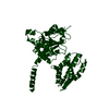
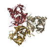
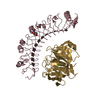
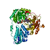
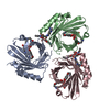
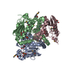
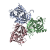
 Vibrio parahaemolyticus (bacteria)
 X-RAY DIFFRACTION /
  Authors
 Citation
 Structure visualization
 Downloads & links
 Other downloads
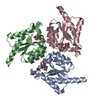


 PDBj
PDBj Assembly
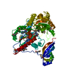

 Mass: 785.550 Da / Num. of mol.: 1 / Source method: obtained synthetically / Formula: C27H33N9O15P2 / Comment: FAD*YM
 Mass: 18.015 Da / Num. of mol.: 112 / Source method: isolated from a natural source / Formula: H2O
 Sample preparation
 / Beamline: X4A / Wavelength: 0.97907 Å
 Processing