 Movie
Movie Controller


+ Open data

- Basic information
| Entry | Database: PDB / ID: 4x9s | ||||||
|---|---|---|---|---|---|---|---|
| Title | CRYSTAL STRUCTURE OF HISAP FROM STREPTOMYCES SP. MG1 | ||||||
 Components Components | Phosphoribosyl isomerase A | ||||||
 Keywords Keywords | ISOMERASE / TIM-BARREL / Structural Genomics / PSI-Biology / Midwest Center for Structural Genomics / MCSG | ||||||
| Function / homology |  Function and homology information Function and homology informationphosphoribosylanthranilate isomerase / phosphoribosylanthranilate isomerase activity / 1-(5-phosphoribosyl)-5-[(5-phosphoribosylamino)methylideneamino]imidazole-4-carboxamide isomerase / 1-(5-phosphoribosyl)-5-[(5-phosphoribosylamino)methylideneamino]imidazole-4-carboxamide isomerase activity / L-histidine biosynthetic process / L-tryptophan biosynthetic process / cytoplasm Similarity search - Function | ||||||
| Biological species |  Streptomyces sp. Mg1 (bacteria) Streptomyces sp. Mg1 (bacteria) | ||||||
| Method |  X-RAY DIFFRACTION / SYNCHROTRON / MOLECULAR REPLACEMENT / Resolution: 1.6 Å X-RAY DIFFRACTION / SYNCHROTRON / MOLECULAR REPLACEMENT / Resolution: 1.6 Å | ||||||
 Authors Authors | MICHALSKA, K. / VERDUZCO-CASTRO, E.A. / ENDRES, M. / BARONA-GOMEZ, F. / JOACHIMIAK, A. / Midwest Center for Structural Genomics (MCSG) | ||||||
| Funding support |  United States, 1items United States, 1items
| ||||||
 Citation Citation | Journal: Biochem. J. / Year: 2016 Title: Co-occurrence of analogous enzymes determines evolution of a novel ( beta alpha )8-isomerase sub-family after non-conserved mutations in flexible loop. Authors: Verduzco-Castro, E.A. / Michalska, K. / Endres, M. / Juarez-Vazquez, A.L. / Noda-Garcia, L. / Chang, C. / Henry, C.S. / Babnigg, G. / Joachimiak, A. / Barona-Gomez, F. | ||||||
| History |
|
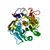
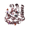
- Structure visualization
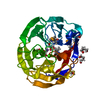
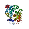
| Structure viewer | Molecule: MolmilJmol/JSmol |
|---|
- Downloads & links
-Download
| PDBx/mmCIF format | 4x9s.cif.gz | 111.6 KB | Display | PDBx/mmCIF format |
|---|---|---|---|---|
| PDB format | pdb4x9s.ent.gz | 83.8 KB | Display | PDB format |
| PDBx/mmJSON format | 4x9s.json.gz | Tree view | PDBx/mmJSON format | |
| Others |  Other downloads Other downloads |
-Validation report
| Arichive directory | https://data.pdbj.org/pub/pdb/validation_reports/x9/4x9sftp://data.pdbj.org/pub/pdb/validation_reports/x9/4x9s | HTTPS FTP |
|---|
-Related structure data
| Related structure data | 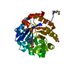 4tx9C 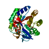 4u28C 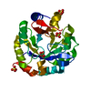 4w9tSC 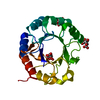 4wuiC 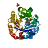 5dn1C S: Starting model for refinement C: citing same article ( |
|---|---|
| Similar structure data | |
| Other databases |


-Links
 PDBj
PDBj
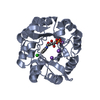
- Assembly
| Deposited unit | 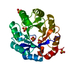
| ||||||||
|---|---|---|---|---|---|---|---|---|---|
| 1 |
| ||||||||
| Unit cell |
|
-Components
| #1: Protein | Mass: 25330.543 Da / Num. of mol.: 1 Source method: isolated from a genetically manipulated source Source: (gene. exp.) Streptomyces sp. Mg1 (bacteria) / Gene: priA, hisA, SSAG_02214 / Plasmid: pMCSG68 / Production host: | ||
|---|---|---|---|
| #2: Chemical | ChemComp-SO4 /   Mass: 96.063 Da / Num. of mol.: 5 / Source method: obtained synthetically / Formula: SO4 Mass: 96.063 Da / Num. of mol.: 5 / Source method: obtained synthetically / Formula: SO4#3: Water | ChemComp-HOH / |  Mass: 18.015 Da / Num. of mol.: 227 / Source method: isolated from a natural source / Formula: H2O Mass: 18.015 Da / Num. of mol.: 227 / Source method: isolated from a natural source / Formula: H2O |
-Experimental details
-Experiment
| Experiment | Method: X-RAY DIFFRACTION / Number of used crystals: 1 |
|---|
- Sample preparation
| Crystal | Density Matthews: 3.04 Å3/Da / Density % sol: 59.53 % |
|---|---|
| Crystal grow | Temperature: 289 K / Method: vapor diffusion, sitting drop Details: 0.1 M Tris/HCl, pH 8.5, 1.5 M Li2SO4 and 2 mM rCdRP PH range: 8.5 |
-Data collection
| Diffraction | Mean temperature: 100 K |
|---|---|
| Diffraction source | Source: SYNCHROTRON / Site: APS / Beamline: 19-ID / Wavelength: 0.97927 Å |
| Detector | Type: ADSC QUANTUM 315r / Detector: CCD / Date: Oct 11, 2013 / Details: mirrors |
| Radiation | Monochromator: SI(111) / Protocol: SINGLE WAVELENGTH / Monochromatic (M) / Laue (L): M / Scattering type: x-ray |
| Radiation wavelength | Wavelength: 0.97927 Å / Relative weight: 1 |
| Reflection | Resolution: 1.6→30 Å / Num. obs: 41756 / % possible obs: 99.2 % / Observed criterion σ(I): -3 / Redundancy: 4.6 % / Rmerge(I) obs: 0.073 / Net I/σ(I): 18.67 |
| Reflection shell | Resolution: 1.6→1.63 Å / Redundancy: 4.1 % / Rmerge(I) obs: 0.638 / Mean I/σ(I) obs: 2 / % possible all: 95.7 |
- Processing
| Software |
| ||||||||||||||||||||||||||||||||||||||||||||||||||||||||||||||||||||||||||||||||||||||||||||||||||||
|---|---|---|---|---|---|---|---|---|---|---|---|---|---|---|---|---|---|---|---|---|---|---|---|---|---|---|---|---|---|---|---|---|---|---|---|---|---|---|---|---|---|---|---|---|---|---|---|---|---|---|---|---|---|---|---|---|---|---|---|---|---|---|---|---|---|---|---|---|---|---|---|---|---|---|---|---|---|---|---|---|---|---|---|---|---|---|---|---|---|---|---|---|---|---|---|---|---|---|---|---|---|
| Refinement | Method to determine structure: MOLECULAR REPLACEMENT Starting model: 4W9T Resolution: 1.6→28.943 Å / SU ML: 0.14 / Cross valid method: THROUGHOUT / σ(F): 0 / Phase error: 18.26 / Stereochemistry target values: ML / Details: HYDROGEN ATOMS HAVE BEEN ADDED IN RIDING POSITIONS
| ||||||||||||||||||||||||||||||||||||||||||||||||||||||||||||||||||||||||||||||||||||||||||||||||||||
| Solvent computation | Shrinkage radii: 0.9 Å / VDW probe radii: 1.11 Å / Solvent model: FLAT BULK SOLVENT MODEL | ||||||||||||||||||||||||||||||||||||||||||||||||||||||||||||||||||||||||||||||||||||||||||||||||||||
| Refinement step | Cycle: LAST / Resolution: 1.6→28.943 Å
| ||||||||||||||||||||||||||||||||||||||||||||||||||||||||||||||||||||||||||||||||||||||||||||||||||||
| Refine LS restraints |
| ||||||||||||||||||||||||||||||||||||||||||||||||||||||||||||||||||||||||||||||||||||||||||||||||||||
| LS refinement shell |
| ||||||||||||||||||||||||||||||||||||||||||||||||||||||||||||||||||||||||||||||||||||||||||||||||||||
| Refinement TLS params. | Method: refined / Refine-ID: X-RAY DIFFRACTION
| ||||||||||||||||||||||||||||||||||||||||||||||||||||||||||||||||||||||||||||||||||||||||||||||||||||
| Refinement TLS group |
|
