 Movie
Movie Controller


+ Open data

- Basic information
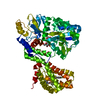
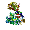
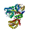
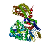
| Entry | Database: PDB / ID: 4wmv | |||||||||
|---|---|---|---|---|---|---|---|---|---|---|
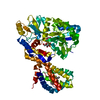
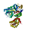
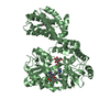
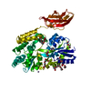
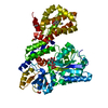
| Title | STRUCTURE OF MBP-MCL1 BOUND TO ligand 4 AT 2.4A | |||||||||
 Components Components | MBL-MCL1 chimera protein | |||||||||
 Keywords Keywords | APOPTOSIS / protein-protein interaction | |||||||||
| Function / homology |  Function and homology information Function and homology informationpositive regulation of oxidative stress-induced neuron intrinsic apoptotic signaling pathway / cell fate determination / cellular homeostasis / mitochondrial fusion / Bcl-2 family protein complex / detection of maltose stimulus / maltose transport complex / carbohydrate transport / BH3 domain binding / negative regulation of anoikis ...positive regulation of oxidative stress-induced neuron intrinsic apoptotic signaling pathway / cell fate determination / cellular homeostasis / mitochondrial fusion / Bcl-2 family protein complex / detection of maltose stimulus / maltose transport complex / carbohydrate transport / BH3 domain binding / negative regulation of anoikis / protein transmembrane transporter activity / negative regulation of extrinsic apoptotic signaling pathway in absence of ligand / carbohydrate transmembrane transporter activity / maltose binding / maltose transport / maltodextrin transmembrane transport / extrinsic apoptotic signaling pathway in absence of ligand / ATP-binding cassette (ABC) transporter complex, substrate-binding subunit-containing / response to cytokine / negative regulation of autophagy / ATP-binding cassette (ABC) transporter complex / release of cytochrome c from mitochondria / cell chemotaxis / intrinsic apoptotic signaling pathway in response to DNA damage / Signaling by ALK fusions and activated point mutants / positive regulation of neuron apoptotic process / outer membrane-bounded periplasmic space / channel activity / Interleukin-4 and Interleukin-13 signaling / regulation of apoptotic process / mitochondrial outer membrane / periplasmic space / positive regulation of apoptotic process / protein heterodimerization activity / DNA damage response / negative regulation of apoptotic process / mitochondrion / nucleoplasm / nucleus / membrane / cytosol / cytoplasm Similarity search - Function | |||||||||
| Biological species |   Homo sapiens (human) Homo sapiens (human) | |||||||||
| Method |  X-RAY DIFFRACTION / MOLECULAR REPLACEMENT / Resolution: 2.4 Å X-RAY DIFFRACTION / MOLECULAR REPLACEMENT / Resolution: 2.4 Å | |||||||||
 Authors Authors | Clifton, M.C. / Moulin, A. | |||||||||
 Citation Citation | Journal: Plos One / Year: 2015 Title: A Maltose-Binding Protein Fusion Construct Yields a Robust Crystallography Platform for MCL1. Authors: Clifton, M.C. / Dranow, D.M. / Leed, A. / Fulroth, B. / Fairman, J.W. / Abendroth, J. / Atkins, K.A. / Wallace, E. / Fan, D. / Xu, G. / Ni, Z.J. / Daniels, D. / Van Drie, J. / Wei, G. / ...Authors: Clifton, M.C. / Dranow, D.M. / Leed, A. / Fulroth, B. / Fairman, J.W. / Abendroth, J. / Atkins, K.A. / Wallace, E. / Fan, D. / Xu, G. / Ni, Z.J. / Daniels, D. / Van Drie, J. / Wei, G. / Burgin, A.B. / Golub, T.R. / Hubbard, B.K. / Serrano-Wu, M.H. | |||||||||
| History |
|
- Structure visualization
| Structure viewer | Molecule: MolmilJmol/JSmol |
|---|
- Downloads & links
-Download
| PDBx/mmCIF format | 4wmv.cif.gz | 213.7 KB | Display | PDBx/mmCIF format |
|---|---|---|---|---|
| PDB format | pdb4wmv.ent.gz | 165.6 KB | Display | PDB format |
| PDBx/mmJSON format | 4wmv.json.gz | Tree view | PDBx/mmJSON format | |
| Others |  Other downloads Other downloads |
-Validation report
| Arichive directory | https://data.pdbj.org/pub/pdb/validation_reports/wm/4wmvftp://data.pdbj.org/pub/pdb/validation_reports/wm/4wmv | HTTPS FTP |
|---|
-Related structure data
| Related structure data | 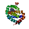 4wmrC 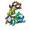 4wmsC  4wmtC  4wmuC  4wmwC 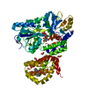 4wmxC  4mbpS  4oq6S C: citing same article ( S: Starting model for refinement |
|---|---|
| Similar structure data |

-Links
 PDBj
PDBj


- Assembly
| Deposited unit | 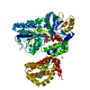
| ||||||||
|---|---|---|---|---|---|---|---|---|---|
| 1 |
| ||||||||
| Unit cell |
|
-Components
-Protein / Sugars , 2 types, 2 molecules A
| #1: Protein | Mass: 57310.883 Da / Num. of mol.: 1 / Mutation: K194A, K197A, R201A Source method: isolated from a genetically manipulated source Source: (gene. exp.) Homo sapiens (human)Strain: K12 / Gene: malE, b4034, JW3994, MCL1, BCL2L3 / Production host: |
|---|---|
| #2: Polysaccharide | alpha-D-glucopyranose-(1-4)-alpha-D-glucopyranose / alpha-maltose  Source method: isolated from a genetically manipulated source Details: oligosaccharide / References: alpha-maltose |
-Non-polymers , 4 types, 148 molecules 


| #3: Chemical | ChemComp-CL /  Mass: 35.453 Da / Num. of mol.: 1 / Source method: obtained synthetically / Formula: Cl Mass: 35.453 Da / Num. of mol.: 1 / Source method: obtained synthetically / Formula: Cl |
|---|---|
| #4: Chemical | ChemComp-MG /  Mass: 24.305 Da / Num. of mol.: 1 / Source method: obtained synthetically / Formula: Mg Mass: 24.305 Da / Num. of mol.: 1 / Source method: obtained synthetically / Formula: Mg |
| #5: Chemical | ChemComp-3R4 /  Mass: 230.643 Da / Num. of mol.: 1 / Source method: obtained synthetically / Formula: C9H4ClFO2S Mass: 230.643 Da / Num. of mol.: 1 / Source method: obtained synthetically / Formula: C9H4ClFO2S |
| #6: Water | ChemComp-HOH / Mass: 18.015 Da / Num. of mol.: 145 / Source method: isolated from a natural source / Formula: H2O |
-Experimental details
-Experiment
| Experiment | Method: X-RAY DIFFRACTION / Number of used crystals: 1 |
|---|
- Sample preparation
| Crystal | Density Matthews: 2.22 Å3/Da / Density % sol: 44.61 % |
|---|---|
| Crystal grow | Temperature: 298 K / Method: vapor diffusion, sitting drop / pH: 7 Details: 10 MG/ML MBP-MCL1, 200MM MG FORMATE, 20% PEG3350, 1MM MALTOSE, CRYOPROTECTANT 20% ethylene glycol, SOAKED IN 10MM ligand for 2 DAYS |
-Data collection
| Diffraction | Mean temperature: 100 K |
|---|---|
| Diffraction source | Source: ROTATING ANODE / Type: RIGAKU FR-E+ SUPERBRIGHT / Wavelength: 1.54 Å |
| Detector | Type: RIGAKU SATURN 944+ / Detector: CCD / Date: May 13, 2014 |
| Radiation | Protocol: SINGLE WAVELENGTH / Monochromatic (M) / Laue (L): M / Scattering type: x-ray |
| Radiation wavelength | Wavelength: 1.54 Å / Relative weight: 1 |
| Reflection | Resolution: 2.4→50 Å / Num. obs: 20683 / % possible obs: 99.6 % / Observed criterion σ(I): -3 / Redundancy: 4.2 % / Biso Wilson estimate: 29.22 Å2 / Rmerge F obs: 0.991 / Rmerge(I) obs: 0.138 / Rrim(I) all: 0.157 / Χ2: 0.901 / Net I/σ(I): 10.33 / Num. measured all: 87886 |
| Reflection shell | Resolution: 2.4→2.46 Å / Rmerge F obs: 0.999 / Rmerge(I) obs: 0.515 / Mean I/σ(I) obs: 3.4 / Num. measured obs: 981 / Num. possible: 289 / Num. unique obs: 272 / Rrim(I) all: 0.034 / Rejects: 0 / % possible all: 99.4 |
- Processing
| Software |
| |||||||||||||||||||||||||||||||||||||||||||||||||||||||||||||||||||||||||||
|---|---|---|---|---|---|---|---|---|---|---|---|---|---|---|---|---|---|---|---|---|---|---|---|---|---|---|---|---|---|---|---|---|---|---|---|---|---|---|---|---|---|---|---|---|---|---|---|---|---|---|---|---|---|---|---|---|---|---|---|---|---|---|---|---|---|---|---|---|---|---|---|---|---|---|---|---|
| Refinement | Method to determine structure: MOLECULAR REPLACEMENT Starting model: 4OQ6,4MBP Resolution: 2.4→40 Å / Cross valid method: FREE R-VALUE / Stereochemistry target values: ML
| |||||||||||||||||||||||||||||||||||||||||||||||||||||||||||||||||||||||||||
| Solvent computation | Shrinkage radii: 0.9 Å / Solvent model: FLAT BULK SOLVENT MODEL | |||||||||||||||||||||||||||||||||||||||||||||||||||||||||||||||||||||||||||
| Displacement parameters | Biso max: 101.64 Å2 / Biso mean: 27.36 Å2 / Biso min: 9.76 Å2 | |||||||||||||||||||||||||||||||||||||||||||||||||||||||||||||||||||||||||||
| Refinement step | Cycle: final / Resolution: 2.4→40 Å
| |||||||||||||||||||||||||||||||||||||||||||||||||||||||||||||||||||||||||||
| Refinement TLS params. | Method: refined / Refine-ID: X-RAY DIFFRACTION
| |||||||||||||||||||||||||||||||||||||||||||||||||||||||||||||||||||||||||||
| Refinement TLS group |
|
