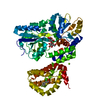
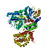
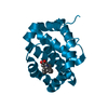
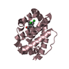
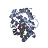
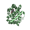
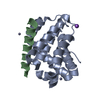
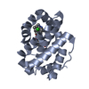
positive regulation of oxidative stress-induced neuron intrinsic apoptotic signaling pathway / cell fate determination / cellular homeostasis / Bcl-2 family protein complex / mitochondrial fusion / negative regulation of anoikis / BH3 domain binding / transmembrane protein transporter activity / negative regulation of extrinsic apoptotic signaling pathway in absence of ligand / extrinsic apoptotic signaling pathway in absence of ligand ...positive regulation of oxidative stress-induced neuron intrinsic apoptotic signaling pathway / cell fate determination / cellular homeostasis / Bcl-2 family protein complex / mitochondrial fusion / negative regulation of anoikis / BH3 domain binding / transmembrane protein transporter activity / negative regulation of extrinsic apoptotic signaling pathway in absence of ligand / extrinsic apoptotic signaling pathway in absence of ligand / response to cytokine / release of cytochrome c from mitochondria / negative regulation of autophagy / intrinsic apoptotic signaling pathway in response to DNA damage / Signaling by ALK fusions and activated point mutants / positive regulation of neuron apoptotic process / channel activity / Interleukin-4 and Interleukin-13 signaling / regulation of apoptotic process / mitochondrial outer membrane / positive regulation of apoptotic process / protein heterodimerization activity / DNA damage response / negative regulation of apoptotic process / mitochondrion / nucleoplasm / membrane / nucleus / cytoplasm / cytosol Similarity search - Function
Apoptosis regulator, Mcl-1 / Blc2-like / Apoptosis Regulator Bcl-x / Apoptosis regulator, Bcl-2, BH3 motif, conserved site / Apoptosis regulator, Bcl-2 family BH3 motif signature. / Apoptosis regulator, Bcl-2, BH1 motif, conserved site / Apoptosis regulator, Bcl-2 family BH1 motif signature. / Apoptosis regulator, Bcl-2, BH2 motif, conserved site / Apoptosis regulator, Bcl-2 family BH2 motif signature. / Bcl-2 family ...Apoptosis regulator, Mcl-1 / Blc2-like / Apoptosis Regulator Bcl-x / Apoptosis regulator, Bcl-2, BH3 motif, conserved site / Apoptosis regulator, Bcl-2 family BH3 motif signature. / Apoptosis regulator, Bcl-2, BH1 motif, conserved site / Apoptosis regulator, Bcl-2 family BH1 motif signature. / Apoptosis regulator, Bcl-2, BH2 motif, conserved site / Apoptosis regulator, Bcl-2 family BH2 motif signature. / Bcl-2 family / BCL (B-Cell lymphoma); contains BH1, BH2 regions / Bcl2-like / Bcl-2, Bcl-2 homology region 1-3 / Apoptosis regulator proteins, Bcl-2 family / BCL2-like apoptosis inhibitors family profile. / Bcl-2-like superfamily / Orthogonal Bundle / Mainly Alpha Similarity search - Domain/homology
Method to determine structure: MOLECULAR REPLACEMENT / Resolution: 1.7→47.17 Å / Cor.coef. Fo:Fc: 0.96 / Cor.coef. Fo:Fc free: 0.934 / SU B: 3.862 / SU ML: 0.07 / Cross valid method: THROUGHOUT / σ(F): 0 / ESU R: 0.123 / ESU R Free: 0.115 / Stereochemistry target values: MAXIMUM LIKELIHOOD Details: U VALUES : WITH TLS ADDED HYDROGENS HAVE BEEN ADDED IN THE RIDING POSITIONS
Rfactor
Num. reflection
% reflection
Selection details
Rfree
0.206
715
5 %
RANDOM
Rwork
0.169
14164
-
-
obs
0.171
14163
97.3 %
-
Solvent computation
Ion probe radii: 0.8 Å / Shrinkage radii: 0.8 Å / VDW probe radii: 1.2 Å / Solvent model: MASK
In the structure databanks used in Yorodumi, some data are registered as the other names, "COVID-19 virus" and "2019-nCoV". Here are the details of the virus and the list of structure data.
Jan 31, 2019. EMDB accession codes are about to change! (news from PDBe EMDB page)
EMDB accession codes are about to change! (news from PDBe EMDB page)
The allocation of 4 digits for EMDB accession codes will soon come to an end. Whilst these codes will remain in use, new EMDB accession codes will include an additional digit and will expand incrementally as the available range of codes is exhausted. The current 4-digit format prefixed with “EMD-” (i.e. EMD-XXXX) will advance to a 5-digit format (i.e. EMD-XXXXX), and so on. It is currently estimated that the 4-digit codes will be depleted around Spring 2019, at which point the 5-digit format will come into force.
The EM Navigator/Yorodumi systems omit the EMD- prefix.
Related info.:Q: What is EMD? / ID/Accession-code notation in Yorodumi/EM Navigator
Yorodumi is a browser for structure data from EMDB, PDB, SASBDB, etc.
This page is also the successor to EM Navigator detail page, and also detail information page/front-end page for Omokage search.
The word "yorodu" (or yorozu) is an old Japanese word meaning "ten thousand". "mi" (miru) is to see.
Related info.:EMDB / PDB / SASBDB / Comparison of 3 databanks / Yorodumi Search / Aug 31, 2016. New EM Navigator & Yorodumi / Yorodumi Papers / Jmol/JSmol / Function and homology information / Changes in new EM Navigator and Yorodumi
 Movie
Movie Controller


 Open data
 Basic information
 Components
 Keywords
 Function and homology information
 Homo sapiens (human)
 X-RAY DIFFRACTION /
  Authors
 Citation
 Structure visualization
 Downloads & links
 Other downloads
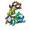


 PDBj
PDBj


 Assembly
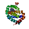


 Mass: 65.409 Da / Num. of mol.: 3 / Source method: obtained synthetically / Formula: Zn
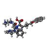
 Mass: 628.720 Da / Num. of mol.: 1 / Source method: obtained synthetically / Formula: C37H36N6O4
 Mass: 175.959 Da / Num. of mol.: 2 / Source method: obtained synthetically / Formula: H2O7P2
 Mass: 18.015 Da / Num. of mol.: 123 / Source method: isolated from a natural source / Formula: H2O
 Sample preparation
 Processing