 Movie
Movie Controller

[English] 日本語
 Yorodumi
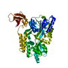
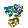
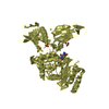
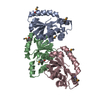
- PDB-3csb: Crystal Structure of Monobody YSX1/Maltose Binding Protein Fusion... -
+ Open data

- Basic information
| Entry | Database: PDB / ID: 3csb | ||||||
|---|---|---|---|---|---|---|---|
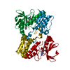
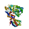
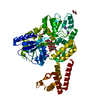
| Title | Crystal Structure of Monobody YSX1/Maltose Binding Protein Fusion Complex | ||||||
 Components Components | Maltose-binding protein Monobody YSX1 Fusion | ||||||
 Keywords Keywords | DE NOVO PROTEIN / SUGAR BINDING PROTEIN / Engineered Binding Protein / Antibody Mimic / Synthetic Protein Interface / Minimalist Protein Interface | ||||||
| Function / homology |  Function and homology information Function and homology informationdetection of maltose stimulus / maltose transport complex / carbohydrate transport / carbohydrate transmembrane transporter activity / maltose binding / maltose transport / maltodextrin transmembrane transport / ATP-binding cassette (ABC) transporter complex, substrate-binding subunit-containing / ATP-binding cassette (ABC) transporter complex / cell chemotaxis ...detection of maltose stimulus / maltose transport complex / carbohydrate transport / carbohydrate transmembrane transporter activity / maltose binding / maltose transport / maltodextrin transmembrane transport / ATP-binding cassette (ABC) transporter complex, substrate-binding subunit-containing / ATP-binding cassette (ABC) transporter complex / cell chemotaxis / outer membrane-bounded periplasmic space / periplasmic space / DNA damage response / membrane Similarity search - Function | ||||||
| Biological species |  synthetic (others) | ||||||
| Method |  X-RAY DIFFRACTION / SYNCHROTRON / MOLECULAR REPLACEMENT / Resolution: 1.999 Å X-RAY DIFFRACTION / SYNCHROTRON / MOLECULAR REPLACEMENT / Resolution: 1.999 Å | ||||||
 Authors Authors | Gilbreth, R.N. / Koide, S. | ||||||
 Citation Citation | Journal: J.Mol.Biol. / Year: 2008 Title: A Dominant Conformational Role for Amino Acid Diversity in Minimalist Protein-Protein Interfaces Authors: Gilbreth, R.N. / Esaki, K. / Koide, A. / Sidhu, S.S. / Koide, S. #1: Journal: Proc.Natl.Acad.Sci.USA / Year: 2007Title: High affinity single-domain binding proteins with a binary code interface Authors: Koide, A. / Gilbreth, R.N. / Esaki, K. / Tereshko, V. / Koide, S. | ||||||
| History |
|
- Structure visualization
| Structure viewer | Molecule: MolmilJmol/JSmol |
|---|
- Downloads & links
-Download
| PDBx/mmCIF format | 3csb.cif.gz | 112.3 KB | Display | PDBx/mmCIF format |
|---|---|---|---|---|
| PDB format | pdb3csb.ent.gz | 84.7 KB | Display | PDB format |
| PDBx/mmJSON format | 3csb.json.gz | Tree view | PDBx/mmJSON format | |
| Others |  Other downloads Other downloads |
-Validation report
| Arichive directory | https://data.pdbj.org/pub/pdb/validation_reports/cs/3csbftp://data.pdbj.org/pub/pdb/validation_reports/cs/3csb | HTTPS FTP |
|---|
-Related structure data


-Links
 PDBj
PDBj


- Assembly
| Deposited unit | 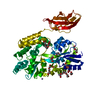
| ||||||||
|---|---|---|---|---|---|---|---|---|---|
| 1 |
| ||||||||
| Unit cell |
|
-Components
-Protein , 1 types, 1 molecules A
| #1: Protein | Mass: 50779.086 Da / Num. of mol.: 1 Source method: isolated from a genetically manipulated source Details: malE includes residues 5-370, Monobody YSX1 377-467, 371-373 comprise an engineered linker Source: (gene. exp.) Escherichia coli, synthetic / Gene: malE / Plasmid: pHFT3 / Production host: |
|---|
-Non-polymers , 7 types, 226 molecules 


| #2: Chemical | ChemComp-MN /  Mass: 54.938 Da / Num. of mol.: 4 / Source method: obtained synthetically / Formula: Mn Mass: 54.938 Da / Num. of mol.: 4 / Source method: obtained synthetically / Formula: Mn#3: Chemical |  Mass: 62.068 Da / Num. of mol.: 2 / Source method: obtained synthetically / Formula: C2H6O2 Mass: 62.068 Da / Num. of mol.: 2 / Source method: obtained synthetically / Formula: C2H6O2#4: Chemical | ChemComp-PGE / |  Mass: 150.173 Da / Num. of mol.: 1 / Source method: obtained synthetically / Formula: C6H14O4 Mass: 150.173 Da / Num. of mol.: 1 / Source method: obtained synthetically / Formula: C6H14O4#5: Chemical | ChemComp-PG4 / |  Mass: 194.226 Da / Num. of mol.: 1 / Source method: obtained synthetically / Formula: C8H18O5 / Comment: precipitant*YM Mass: 194.226 Da / Num. of mol.: 1 / Source method: obtained synthetically / Formula: C8H18O5 / Comment: precipitant*YM#6: Chemical | ChemComp-1PE / |  Mass: 238.278 Da / Num. of mol.: 1 / Source method: obtained synthetically / Formula: C10H22O6 / Comment: precipitant*YM Mass: 238.278 Da / Num. of mol.: 1 / Source method: obtained synthetically / Formula: C10H22O6 / Comment: precipitant*YM#7: Chemical | ChemComp-PEG /  Mass: 106.120 Da / Num. of mol.: 9 / Source method: obtained synthetically / Formula: C4H10O3 Mass: 106.120 Da / Num. of mol.: 9 / Source method: obtained synthetically / Formula: C4H10O3#8: Water | ChemComp-HOH / | Mass: 18.015 Da / Num. of mol.: 208 / Source method: isolated from a natural source / Formula: H2O |
|---|
-Experimental details
-Experiment
| Experiment | Method: X-RAY DIFFRACTION / Number of used crystals: 1 |
|---|
- Sample preparation
| Crystal | Density Matthews: 3.24 Å3/Da / Density % sol: 61.99 % |
|---|---|
| Crystal grow | Temperature: 292 K / Method: vapor diffusion, hanging drop / pH: 6.5 Details: 41% polyethelyeneglycol-400, 2% 2-methyl-2,4-pentanediol, 50 mM MnCl2, 0.1 M 2-(N-morpholino)ethanesulfonic acid, pH 6.5, VAPOR DIFFUSION, HANGING DROP, temperature 292K |
-Data collection
| Diffraction | Mean temperature: 100 K |
|---|---|
| Diffraction source | Source: SYNCHROTRON / Site: APS  / Beamline: 23-ID-D / Wavelength: 1 Å / Beamline: 23-ID-D / Wavelength: 1 Å |
| Detector | Type: MARMOSAIC 300 mm CCD / Detector: CCD / Date: Jul 21, 2007 |
| Radiation | Monochromator: double crystal monochromator and K-B pair of biomorph mirrors for vertical and horizontal focusing Protocol: SINGLE WAVELENGTH / Monochromatic (M) / Laue (L): M / Scattering type: x-ray |
| Radiation wavelength | Wavelength: 1 Å / Relative weight: 1 |
| Reflection | Resolution: 1.999→50 Å / Num. all: 45823 / Num. obs: 45823 / % possible obs: 100 % / Observed criterion σ(F): 0 / Observed criterion σ(I): 0 |
| Reflection shell | Resolution: 1.999→2.07 Å / % possible all: 100 |
- Processing
| Software |
| ||||||||||||||||||||||||||||||||||||||||||||||||||||||||||||||||||||||||||||||||||||||||||
|---|---|---|---|---|---|---|---|---|---|---|---|---|---|---|---|---|---|---|---|---|---|---|---|---|---|---|---|---|---|---|---|---|---|---|---|---|---|---|---|---|---|---|---|---|---|---|---|---|---|---|---|---|---|---|---|---|---|---|---|---|---|---|---|---|---|---|---|---|---|---|---|---|---|---|---|---|---|---|---|---|---|---|---|---|---|---|---|---|---|---|---|
| Refinement | Method to determine structure: MOLECULAR REPLACEMENT / Resolution: 1.999→20 Å / Cor.coef. Fo:Fc: 0.95 / Cor.coef. Fo:Fc free: 0.927 / SU B: 6.123 / SU ML: 0.089 / Cross valid method: THROUGHOUT / σ(F): 0 / ESU R: 0.159 / ESU R Free: 0.151 / Stereochemistry target values: MAXIMUM LIKELIHOOD
| ||||||||||||||||||||||||||||||||||||||||||||||||||||||||||||||||||||||||||||||||||||||||||
| Solvent computation | Ion probe radii: 0.8 Å / Shrinkage radii: 0.8 Å / VDW probe radii: 1.4 Å / Solvent model: BABINET MODEL WITH MASK | ||||||||||||||||||||||||||||||||||||||||||||||||||||||||||||||||||||||||||||||||||||||||||
| Displacement parameters | Biso mean: 37.551 Å2
| ||||||||||||||||||||||||||||||||||||||||||||||||||||||||||||||||||||||||||||||||||||||||||
| Refinement step | Cycle: LAST / Resolution: 1.999→20 Å
| ||||||||||||||||||||||||||||||||||||||||||||||||||||||||||||||||||||||||||||||||||||||||||
| Refine LS restraints |
| ||||||||||||||||||||||||||||||||||||||||||||||||||||||||||||||||||||||||||||||||||||||||||
| LS refinement shell | Resolution: 1.999→2.051 Å / Total num. of bins used: 20
|
