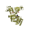
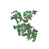
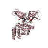
Entry Database : PDB / ID : 3zczTitle Crystal structure of a complex between Actinomadura R39 DD-peptidase and a trifluoroketone inhibitor D-ALANYL-D-ALANINE CARBOXYPEPTIDASE Keywords / / Function / homology Function Domain/homology Component
/ / / / / / / / / / / / / / / / Biological species ACTINOMADURA SP. R39 (bacteria)Method / / / Resolution : 2.6 Å Authors Sauvage, E. / Herman, R. / Kerff, F. / Rocaboy, M. / Charlier, P. Journal : Biochemistry / Year : 2013Title : Inhibition of Dd-Peptidases by a Specific Trifluoroketone: Crystal Structure of a Complex with the Actinomadura R39 Dd-Peptidase.Authors : Dzhekieva, L. / Adediran, S.A. / Herman, R. / Kerff, F. / Duez, C. / Charlier, P. / Sauvage, E. / Pratt, R.F. History Deposition Nov 23, 2012 Deposition site / Processing site Revision 1.0 Mar 27, 2013 Provider / Type Revision 1.1 Aug 28, 2013 Group Revision 1.2 Dec 20, 2023 Group Data collection / Database references ... Data collection / Database references / Derived calculations / Other / Refinement description Category chem_comp_atom / chem_comp_bond ... chem_comp_atom / chem_comp_bond / database_2 / pdbx_database_status / pdbx_initial_refinement_model / pdbx_struct_conn_angle / struct_conn / struct_site Item _database_2.pdbx_DOI / _database_2.pdbx_database_accession ... _database_2.pdbx_DOI / _database_2.pdbx_database_accession / _pdbx_database_status.status_code_sf / _pdbx_struct_conn_angle.ptnr1_auth_comp_id / _pdbx_struct_conn_angle.ptnr1_auth_seq_id / _pdbx_struct_conn_angle.ptnr1_label_asym_id / _pdbx_struct_conn_angle.ptnr1_label_atom_id / _pdbx_struct_conn_angle.ptnr1_label_comp_id / _pdbx_struct_conn_angle.ptnr1_label_seq_id / _pdbx_struct_conn_angle.ptnr3_auth_comp_id / _pdbx_struct_conn_angle.ptnr3_auth_seq_id / _pdbx_struct_conn_angle.ptnr3_label_asym_id / _pdbx_struct_conn_angle.ptnr3_label_atom_id / _pdbx_struct_conn_angle.ptnr3_label_comp_id / _pdbx_struct_conn_angle.ptnr3_label_seq_id / _pdbx_struct_conn_angle.value / _struct_conn.conn_type_id / _struct_conn.id / _struct_conn.pdbx_dist_value / _struct_conn.pdbx_leaving_atom_flag / _struct_conn.ptnr1_auth_asym_id / _struct_conn.ptnr1_auth_comp_id / _struct_conn.ptnr1_auth_seq_id / _struct_conn.ptnr1_label_asym_id / _struct_conn.ptnr1_label_atom_id / _struct_conn.ptnr1_label_comp_id / _struct_conn.ptnr1_label_seq_id / _struct_conn.ptnr2_auth_asym_id / _struct_conn.ptnr2_auth_comp_id / _struct_conn.ptnr2_auth_seq_id / _struct_conn.ptnr2_label_asym_id / _struct_conn.ptnr2_label_atom_id / _struct_conn.ptnr2_label_comp_id / _struct_conn.ptnr2_label_seq_id / _struct_site.pdbx_auth_asym_id / _struct_site.pdbx_auth_comp_id / _struct_site.pdbx_auth_seq_id Revision 1.3 Nov 13, 2024 Group / Category / pdbx_modification_feature
Show all Show less
 Movie
Movie Controller

 Yorodumi
 Open data
 Basic information
 Components
 Keywords
 Function and homology information
 ACTINOMADURA SP. R39 (bacteria)
 X-RAY DIFFRACTION /
  Authors
 Citation
 Structure visualization
 Downloads & links
 Other downloads


 PDBj
PDBj
 Assembly
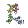

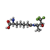
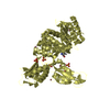
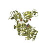
 Type: D-peptide linking / Mass: 300.275 Da / Num. of mol.: 4 / Source method: obtained synthetically / Formula: C11H19F3N2O4
 Mass: 96.063 Da / Num. of mol.: 18 / Source method: obtained synthetically / Formula: SO4
 Mass: 24.305 Da / Num. of mol.: 4 / Source method: obtained synthetically / Formula: Mg
 Mass: 18.015 Da / Num. of mol.: 323 / Source method: isolated from a natural source / Formula: H2O
 Sample preparation
 / Beamline: PROXIMA 1 / Wavelength: 0.9801
  Processing