 Movie
Movie Controller

[English] 日本語
 Yorodumi
- PDB-2y4a: Unexpected tricovalent binding mode of boronic acids within the a... -
+ Open data

- Basic information
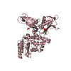
| Entry | Database: PDB / ID: 2y4a | ||||||
|---|---|---|---|---|---|---|---|
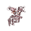
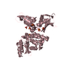
| Title | Unexpected tricovalent binding mode of boronic acids within the active site of a penicillin binding protein | ||||||
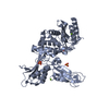
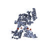
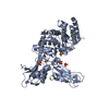
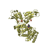
 Components Components | D-ALANYL-D-ALANINE CARBOXYPEPTIDASE | ||||||
 Keywords Keywords | HYDROLASE / PEPTIDOGLYCAN / TETRAVALENT BORON | ||||||
| Function / homology |  Function and homology information Function and homology informationserine-type D-Ala-D-Ala carboxypeptidase / serine-type D-Ala-D-Ala carboxypeptidase activity / peptidoglycan biosynthetic process / cell wall organization / regulation of cell shape / response to antibiotic / proteolysis / extracellular region Similarity search - Function | ||||||
| Biological species |  ACTINOMADURA SP (bacteria) ACTINOMADURA SP (bacteria) | ||||||
| Method |  X-RAY DIFFRACTION / SYNCHROTRON / MOLECULAR REPLACEMENT / Resolution: 2.7 Å X-RAY DIFFRACTION / SYNCHROTRON / MOLECULAR REPLACEMENT / Resolution: 2.7 Å | ||||||
 Authors Authors | Sauvage, E. / Zervosen, A. / Herman, R. / Kerff, F. / Rocaboy, M. / Charlier, P. | ||||||
 Citation Citation | Journal: J.Am.Chem.Soc. / Year: 2011 Title: Unexpected Tricovalent Binding Mode of Boronic Acids within the Active Site of a Penicillin- Binding Protein. Authors: Zervosen, A. / Herman, R. / Kerff, F. / Herman, A. / Bouillez, A. / Prati, F. / Pratt, R.F. / Frere, J.M. / Joris, B. / Luxen, A. / Charlier, P. / Sauvage, E. | ||||||
| History |
|
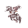
- Structure visualization
| Structure viewer | Molecule: MolmilJmol/JSmol |
|---|
- Downloads & links
-Download
| PDBx/mmCIF format | 2y4a.cif.gz | 667.6 KB | Display | PDBx/mmCIF format |
|---|---|---|---|---|
| PDB format | pdb2y4a.ent.gz | 558.3 KB | Display | PDB format |
| PDBx/mmJSON format | 2y4a.json.gz | Tree view | PDBx/mmJSON format | |
| Others |  Other downloads Other downloads |
-Validation report
| Arichive directory | https://data.pdbj.org/pub/pdb/validation_reports/y4/2y4aftp://data.pdbj.org/pub/pdb/validation_reports/y4/2y4a | HTTPS FTP |
|---|
-Related structure data
| Related structure data |  2y55C  2y59C  3zvtC  3zvwC  2xdmS S: Starting model for refinement C: citing same article ( |
|---|---|
| Similar structure data |


-Links
 PDBj
PDBj
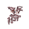
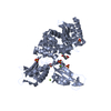
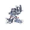
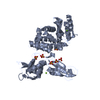
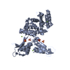
- Assembly
| Deposited unit | 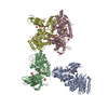
| ||||||||
|---|---|---|---|---|---|---|---|---|---|
| 1 | 
| ||||||||
| 2 | 
| ||||||||
| 3 | 
| ||||||||
| 4 | 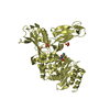
| ||||||||
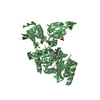
| Unit cell |
|
-Components
| #1: Protein | Mass: 47647.004 Da / Num. of mol.: 4 / Fragment: RESIDUES 50-515 / Source method: isolated from a natural source / Source: (natural) ACTINOMADURA SP (bacteria) / Strain: R39References: UniProt: P39045, serine-type D-Ala-D-Ala carboxypeptidase #2: Chemical | ChemComp-BH6 / {[( 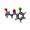  Mass: 213.426 Da / Num. of mol.: 4 / Source method: obtained synthetically / Formula: C8H9BClNO3 Mass: 213.426 Da / Num. of mol.: 4 / Source method: obtained synthetically / Formula: C8H9BClNO3#3: Chemical | ChemComp-SO4 /   Mass: 96.063 Da / Num. of mol.: 20 / Source method: obtained synthetically / Formula: SO4 Mass: 96.063 Da / Num. of mol.: 20 / Source method: obtained synthetically / Formula: SO4#4: Chemical | ChemComp-MG /   Mass: 24.305 Da / Num. of mol.: 4 / Source method: obtained synthetically / Formula: Mg Mass: 24.305 Da / Num. of mol.: 4 / Source method: obtained synthetically / Formula: Mg#5: Water | ChemComp-HOH / |  Mass: 18.015 Da / Num. of mol.: 162 / Source method: isolated from a natural source / Formula: H2O Mass: 18.015 Da / Num. of mol.: 162 / Source method: isolated from a natural source / Formula: H2OHas protein modification | Y | |
|---|
-Experimental details
-Experiment
| Experiment | Method: X-RAY DIFFRACTION / Number of used crystals: 1 |
|---|
- Sample preparation
| Crystal | Density Matthews: 2.55 Å3/Da / Density % sol: 51.5 % / Description: NONE |
|---|---|
| Crystal grow | pH: 6 / Details: pH 6 |
-Data collection
| Diffraction | Mean temperature: 100 K |
|---|---|
| Diffraction source | Source: SYNCHROTRON / Site: ESRF  / Beamline: BM30A / Wavelength: 1.0724 / Beamline: BM30A / Wavelength: 1.0724 |
| Detector | Date: Jan 31, 2009 |
| Radiation | Protocol: SINGLE WAVELENGTH / Monochromatic (M) / Laue (L): M / Scattering type: x-ray |
| Radiation wavelength | Wavelength: 1.0724 Å / Relative weight: 1 |
| Reflection | Resolution: 2.7→46.1 Å / Num. obs: 53645 / % possible obs: 97.7 % / Observed criterion σ(I): 0 / Redundancy: 5.4 % / Rmerge(I) obs: 0.11 / Net I/σ(I): 15.1 |
| Reflection shell | Resolution: 2.7→2.85 Å / Redundancy: 5.4 % / Rmerge(I) obs: 0.57 / Mean I/σ(I) obs: 2.8 / % possible all: 97.1 |
- Processing
| Software |
| ||||||||||||||||||||||||||||||||||||||||||||||||||||||||||||||||||||||||||||||||||||||||||||||||||||||||||||||||||||||||||||||||||||||||||||||||||||||||||||||||||||||||||||||||||||||
|---|---|---|---|---|---|---|---|---|---|---|---|---|---|---|---|---|---|---|---|---|---|---|---|---|---|---|---|---|---|---|---|---|---|---|---|---|---|---|---|---|---|---|---|---|---|---|---|---|---|---|---|---|---|---|---|---|---|---|---|---|---|---|---|---|---|---|---|---|---|---|---|---|---|---|---|---|---|---|---|---|---|---|---|---|---|---|---|---|---|---|---|---|---|---|---|---|---|---|---|---|---|---|---|---|---|---|---|---|---|---|---|---|---|---|---|---|---|---|---|---|---|---|---|---|---|---|---|---|---|---|---|---|---|---|---|---|---|---|---|---|---|---|---|---|---|---|---|---|---|---|---|---|---|---|---|---|---|---|---|---|---|---|---|---|---|---|---|---|---|---|---|---|---|---|---|---|---|---|---|---|---|---|---|
| Refinement | Method to determine structure: MOLECULAR REPLACEMENT Starting model: PDB ENTRY 2XDM Resolution: 2.7→46.1 Å / Cor.coef. Fo:Fc: 0.941 / Cor.coef. Fo:Fc free: 0.911 / SU B: 25.52 / SU ML: 0.294 / Cross valid method: THROUGHOUT / σ(F): 0 / ESU R Free: 0.324 / Stereochemistry target values: MAXIMUM LIKELIHOOD / Details: HYDROGENS HAVE BEEN ADDED IN THE RIDING POSITIONS.
| ||||||||||||||||||||||||||||||||||||||||||||||||||||||||||||||||||||||||||||||||||||||||||||||||||||||||||||||||||||||||||||||||||||||||||||||||||||||||||||||||||||||||||||||||||||||
| Solvent computation | Ion probe radii: 0.8 Å / Shrinkage radii: 0.8 Å / VDW probe radii: 1.4 Å / Solvent model: MASK | ||||||||||||||||||||||||||||||||||||||||||||||||||||||||||||||||||||||||||||||||||||||||||||||||||||||||||||||||||||||||||||||||||||||||||||||||||||||||||||||||||||||||||||||||||||||
| Displacement parameters | Biso mean: 47.5 Å2
| ||||||||||||||||||||||||||||||||||||||||||||||||||||||||||||||||||||||||||||||||||||||||||||||||||||||||||||||||||||||||||||||||||||||||||||||||||||||||||||||||||||||||||||||||||||||
| Refinement step | Cycle: LAST / Resolution: 2.7→46.1 Å
| ||||||||||||||||||||||||||||||||||||||||||||||||||||||||||||||||||||||||||||||||||||||||||||||||||||||||||||||||||||||||||||||||||||||||||||||||||||||||||||||||||||||||||||||||||||||
| Refine LS restraints |
|