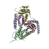
[English] 日本語
Yorodumi
- PDB-3vd8: Crystal structure of human AIM2 PYD domain with MBP fusion -

+
Open data


ID or keywords:

Loading...

-
Basic information

Entry
Database: PDB / ID: 3vd8
TitleCrystal structure of human AIM2 PYD domain with MBP fusion
ComponentsMaltose-binding periplasmic protein, Interferon-inducible protein AIM2
KeywordsSUGAR BINDING PROTEIN / SIGNALING PROTEIN / MBP/PYD/DD / signal transduction / inflammasome
Function / homology
Function and homology information


pyroptosome complex assembly / AIM2 inflammasome complex assembly / The AIM2 inflammasome / cysteine-type endopeptidase activator activity / regulation of behavior / AIM2 inflammasome complex / Cytosolic sensors of pathogen-associated DNA / positive regulation of cysteine-type endopeptidase activity / pattern recognition receptor signaling pathway / pattern recognition receptor activity ...pyroptosome complex assembly / AIM2 inflammasome complex assembly / The AIM2 inflammasome / cysteine-type endopeptidase activator activity / regulation of behavior / AIM2 inflammasome complex / Cytosolic sensors of pathogen-associated DNA / positive regulation of cysteine-type endopeptidase activity / pattern recognition receptor signaling pathway / pattern recognition receptor activity / negative regulation of NF-kappaB transcription factor activity / pyroptotic inflammatory response / T cell homeostasis / carbohydrate transmembrane transporter activity / maltose binding / maltose transport / maltodextrin transmembrane transport / cellular response to interferon-beta / negative regulation of phosphatidylinositol 3-kinase/protein kinase B signal transduction / signaling adaptor activity / ATP-binding cassette (ABC) transporter complex, substrate-binding subunit-containing / positive regulation of defense response to virus by host / tumor necrosis factor-mediated signaling pathway / activation of innate immune response / positive regulation of interleukin-1 beta production / brain development / neuron cellular homeostasis / positive regulation of inflammatory response / cellular response to xenobiotic stimulus / site of double-strand break / positive regulation of NF-kappaB transcription factor activity / outer membrane-bounded periplasmic space / double-stranded DNA binding / defense response to virus / inflammatory response / immune response / innate immune response / DNA damage response / mitochondrion / nucleoplasm / identical protein binding / cytoplasm / cytosol
Similarity search - Function
HIN-200/IF120x / HIN-200 family / HIN-200/IF120x domain / HIN-200 A and B domains profile. / DAPIN domain / DAPIN domain profile. / PAAD/DAPIN/Pyrin domain / Death Domain, Fas / PAAD/DAPIN/Pyrin domain / Death Domain, Fas ...HIN-200/IF120x / HIN-200 family / HIN-200/IF120x domain / HIN-200 A and B domains profile. / DAPIN domain / DAPIN domain profile. / PAAD/DAPIN/Pyrin domain / Death Domain, Fas / PAAD/DAPIN/Pyrin domain / Death Domain, Fas / Maltose/Cyclodextrin ABC transporter, substrate-binding protein / Solute-binding family 1, conserved site / Bacterial extracellular solute-binding proteins, family 1 signature. / Bacterial extracellular solute-binding protein / Bacterial extracellular solute-binding protein / Death-like domain superfamily / Periplasmic binding protein-like II / D-Maltodextrin-Binding Protein; domain 2 / Nucleic acid-binding, OB-fold / Orthogonal Bundle / 3-Layer(aba) Sandwich / Mainly Alpha / Alpha Beta
Similarity search - Domain/homology
alpha-maltotetraose / : / Interferon-inducible protein AIM2 / Maltose/maltodextrin-binding periplasmic protein
Similarity search - Component
Biological speciesEscherichia coli (E. coli)
Homo sapiens (human)
MethodX-RAY DIFFRACTION / SYNCHROTRON / MOLECULAR REPLACEMENT / Resolution: 2.0685 Å
AuthorsJin, T.C. / Perry, A. / Smith, P. / Xiao, T.S.
CitationJournal: J.Biol.Chem. / Year: 2013
Title: Structure of the Absent in Melanoma 2 (AIM2) Pyrin Domain Provides Insights into the Mechanisms of AIM2 Autoinhibition and Inflammasome Assembly.
Authors: Jin, T. / Perry, A. / Smith, P. / Jiang, J. / Xiao, T.S.
History
DepositionJan 4, 2012Deposition site: RCSB / Processing site: RCSB
Revision 1.0Jan 16, 2013Provider: repository / Type: Initial release
Revision 1.1Feb 13, 2013Group: Database references / Structure summary
Revision 1.2Jun 5, 2013Group: Database references
Revision 1.3Aug 2, 2017Group: Refinement description / Source and taxonomy / Category: entity_src_gen / software
Revision 2.0Jul 29, 2020Group: Atomic model / Data collection ...Atomic model / Data collection / Database references / Derived calculations / Non-polymer description / Structure summary
Category: atom_site / chem_comp ...atom_site / chem_comp / entity / entity_name_com / pdbx_branch_scheme / pdbx_chem_comp_identifier / pdbx_entity_branch / pdbx_entity_branch_descriptor / pdbx_entity_branch_link / pdbx_entity_branch_list / pdbx_entity_nonpoly / pdbx_molecule_features / pdbx_nonpoly_scheme / pdbx_struct_conn_angle / struct_asym / struct_conn / struct_conn_type / struct_ref_seq_dif / struct_site / struct_site_gen
Item: _atom_site.B_iso_or_equiv / _atom_site.Cartn_x ..._atom_site.B_iso_or_equiv / _atom_site.Cartn_x / _atom_site.Cartn_y / _atom_site.Cartn_z / _atom_site.auth_asym_id / _atom_site.auth_atom_id / _atom_site.auth_comp_id / _atom_site.auth_seq_id / _atom_site.label_asym_id / _atom_site.label_atom_id / _atom_site.label_comp_id / _atom_site.label_entity_id / _atom_site.type_symbol / _chem_comp.formula / _chem_comp.formula_weight / _chem_comp.id / _chem_comp.mon_nstd_flag / _chem_comp.name / _chem_comp.type / _entity.formula_weight / _entity.pdbx_description / _entity.pdbx_number_of_molecules / _entity.src_method / _entity.type / _pdbx_struct_conn_angle.ptnr1_auth_comp_id / _pdbx_struct_conn_angle.ptnr1_auth_seq_id / _pdbx_struct_conn_angle.ptnr1_label_asym_id / _pdbx_struct_conn_angle.ptnr1_label_atom_id / _pdbx_struct_conn_angle.ptnr1_label_comp_id / _pdbx_struct_conn_angle.ptnr1_label_seq_id / _pdbx_struct_conn_angle.ptnr2_auth_seq_id / _pdbx_struct_conn_angle.ptnr2_label_asym_id / _pdbx_struct_conn_angle.ptnr3_auth_comp_id / _pdbx_struct_conn_angle.ptnr3_auth_seq_id / _pdbx_struct_conn_angle.ptnr3_label_asym_id / _pdbx_struct_conn_angle.ptnr3_label_atom_id / _pdbx_struct_conn_angle.ptnr3_label_comp_id / _pdbx_struct_conn_angle.ptnr3_label_seq_id / _pdbx_struct_conn_angle.value / _struct_asym.entity_id / _struct_ref_seq_dif.details
Description: Carbohydrate remediation / Provider: repository / Type: Remediation
Revision 2.1Sep 13, 2023Group: Data collection / Database references ...Data collection / Database references / Refinement description / Structure summary
Category: chem_comp / chem_comp_atom ...chem_comp / chem_comp_atom / chem_comp_bond / database_2 / pdbx_initial_refinement_model
Item: _chem_comp.pdbx_synonyms / _database_2.pdbx_DOI / _database_2.pdbx_database_accession

-
Structure visualization

Structure viewerMolecule:
MolmilJmol/JSmol

Downloads & links

-
Assembly

Deposited unit
A: Maltose-binding periplasmic protein, Interferon-inducible protein AIM2
hetero molecules


Theoretical massNumber of molelcules
Total (without water)55,04510
Polymers53,9731
Non-polymers1,0719
Water5,855325
1


  • Idetical with deposited unit
  • defined by author&software
TypeNameSymmetry operationNumber
identity operation1_555x,y,z1
MethodPISA
Unit cell
Length a, b, c (Å)61.599, 91.934, 100.281
Angle α, β, γ (deg.)90.00, 90.00, 90.00
Int Tables number19
Space group name H-MP212121

-
Components

#1: Protein Maltose-binding periplasmic protein, Interferon-inducible protein AIM2 / MBP / MMBP / Maltodextrin-binding protein / Absent in melanoma 2


Mass: 53973.414 Da / Num. of mol.: 1 / Fragment: PYD domain (UNP Residues 1-107) / Mutation: D82A, K83A, E172A, N173A, K239A
Source method: isolated from a genetically manipulated source
Source: (gene. exp.) Escherichia coli (E. coli), (gene. exp.) Homo sapiens (human)
Strain: O157:H7 / Gene: AIM2, ECs5017, malE, MBP, Z5632 / Plasmid: pET30a / Production host: Escherichia coli (E. coli) / Strain (production host): BL21(DE3) / References: UniProt: P0AEY0, UniProt: O14862
#2: Polysaccharide alpha-D-glucopyranose-(1-4)-alpha-D-glucopyranose-(1-4)-alpha-D-glucopyranose-(1-4)-alpha-D-glucopyranose / alpha-maltotetraose


Type: oligosaccharide, Oligosaccharide / Class: Substrate analog / Mass: 666.578 Da / Num. of mol.: 1
Source method: isolated from a genetically manipulated source
Details: oligosaccharide / References: alpha-maltotetraose
DescriptorTypeProgram
DGlcpa1-4DGlcpa1-4DGlcpa1-4DGlcpa1-ROHGlycam Condensed SequenceGMML 1.0
WURCS=2.0/1,4,3/[a2122h-1a_1-5]/1-1-1-1/a4-b1_b4-c1_c4-d1WURCSPDB2Glycan 1.1.0
[][a-D-Glcp]{[(4+1)][a-D-Glcp]{[(4+1)][a-D-Glcp]{[(4+1)][a-D-Glcp]{}}}}LINUCSPDB-CARE
#3: Chemical
ChemComp-EDO / 1,2-ETHANEDIOL / ETHYLENE GLYCOL


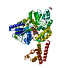
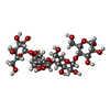
Mass: 62.068 Da / Num. of mol.: 4 / Source method: obtained synthetically / Formula: C2H6O2
#4: Chemical
ChemComp-K / POTASSIUM ION


Mass: 39.098 Da / Num. of mol.: 4 / Source method: obtained synthetically / Formula: K
#5: Water ChemComp-HOH / water


Mass: 18.015 Da / Num. of mol.: 325 / Source method: isolated from a natural source / Formula: H2O

-
Experimental details

-
Experiment

ExperimentMethod: X-RAY DIFFRACTION / Number of used crystals: 1

-
Sample preparation

CrystalDensity Matthews: 2.63 Å3/Da / Density % sol: 53.24 %
Crystal growTemperature: 298 K / Method: vapor diffusion, hanging drop / pH: 4
Details: 20% PEG3350, 0.1M Potassium Acetate, pH 4.0, VAPOR DIFFUSION, HANGING DROP, temperature 298K

-
Data collection

DiffractionMean temperature: 100 K
Diffraction sourceSource: SYNCHROTRON / Site: APS / Beamline: 23-ID-B / Wavelength: 0.9794 Å
DetectorType: MARMOSAIC 300 mm CCD / Detector: CCD / Date: Mar 7, 2011 / Details: mirrors
RadiationMonochromator: Si 111 CHANNEL / Protocol: SINGLE WAVELENGTH / Monochromatic (M) / Laue (L): M / Scattering type: x-ray
Radiation wavelengthWavelength: 0.9794 Å / Relative weight: 1
ReflectionResolution: 2.068→50 Å / Num. all: 35519 / Num. obs: 35164 / % possible obs: 99 % / Observed criterion σ(F): 0 / Observed criterion σ(I): 0 / Redundancy: 7 % / Net I/σ(I): 12.36
Reflection shellResolution: 2.068→2.11 Å / Redundancy: 6 % / Rmerge(I) obs: 0.646 / Mean I/σ(I) obs: 2.24 / Num. unique all: 1704 / % possible all: 97

-
Processing

Software
NameVersionClassification
Blu-Icedata collection
PHASERphasing
PHENIX(phenix.refine: dev_947)refinement
HKL-2000data reduction
HKL-2000data scaling
RefinementMethod to determine structure: MOLECULAR REPLACEMENT
Starting model: PDB ENTRY 3OB4
Resolution: 2.0685→38.887 Å / SU ML: 0.21 / σ(F): 0.82 / Phase error: 21.38 / Stereochemistry target values: ML
RfactorNum. reflection% reflectionSelection details
Rfree0.2212 1741 4.99 %random
Rwork0.1821 ---
obs0.1841 34906 98.33 %-
all-35519 --
Solvent computationShrinkage radii: 0.9 Å / VDW probe radii: 1.11 Å / Solvent model: FLAT BULK SOLVENT MODEL / Bsol: 54.696 Å2 / ksol: 0.353 e/Å3
Displacement parameters
Baniso -1Baniso -2Baniso -3
1--0.233 Å20 Å20 Å2
2--0.1537 Å20 Å2
3---0.0793 Å2
Refinement stepCycle: LAST / Resolution: 2.0685→38.887 Å
ProteinNucleic acidLigandSolventTotal
Num. atoms3630 0 65 325 4020
Refine LS restraints
Refine-IDTypeDev idealNumber
X-RAY DIFFRACTIONf_bond_d0.0033832
X-RAY DIFFRACTIONf_angle_d0.6945197
X-RAY DIFFRACTIONf_dihedral_angle_d11.3911400
X-RAY DIFFRACTIONf_chiral_restr0.05585
X-RAY DIFFRACTIONf_plane_restr0.003663
LS refinement shell
Resolution (Å)Rfactor RfreeNum. reflection RfreeRfactor RworkNum. reflection RworkRefine-ID% reflection obs (%)
2.0685-2.12930.28121200.24572713X-RAY DIFFRACTION97
2.1293-2.1980.28381430.21872693X-RAY DIFFRACTION98
2.198-2.27660.25591390.20342713X-RAY DIFFRACTION98
2.2766-2.36770.2521500.19362741X-RAY DIFFRACTION98
2.3677-2.47550.23161270.18992753X-RAY DIFFRACTION99
2.4755-2.6060.23441530.19612757X-RAY DIFFRACTION99
2.606-2.76920.24041430.19812762X-RAY DIFFRACTION99
2.7692-2.98290.24461580.19632759X-RAY DIFFRACTION99
2.9829-3.2830.24461530.19362811X-RAY DIFFRACTION100
3.283-3.75770.19781410.15832809X-RAY DIFFRACTION100
3.7577-4.7330.18011700.14992838X-RAY DIFFRACTION100
4.733-38.89340.21871440.18732816X-RAY DIFFRACTION94

+
About Yorodumi

-
News

-
Feb 9, 2022. New format data for meta-information of EMDB entries

New format data for meta-information of EMDB entries

  • Version 3 of the EMDB header file is now the official format.
  • The previous official version 1.9 will be removed from the archive.

Related info.:EMDB header

External links:wwPDB to switch to version 3 of the EMDB data model

-
Aug 12, 2020. Covid-19 info

Covid-19 info

URL: https://pdbjlvh1.pdbj.org/emnavi/covid19.php

New page: Covid-19 featured information page in EM Navigator.

Related info.:Covid-19 info / Mar 5, 2020. Novel coronavirus structure data

+
Mar 5, 2020. Novel coronavirus structure data

Novel coronavirus structure data

Related info.:Yorodumi Speices / Aug 12, 2020. Covid-19 info

External links:COVID-19 featured content - PDBj / Molecule of the Month (242):Coronavirus Proteases

+
Jan 31, 2019. EMDB accession codes are about to change! (news from PDBe EMDB page)

EMDB accession codes are about to change! (news from PDBe EMDB page)

  • The allocation of 4 digits for EMDB accession codes will soon come to an end. Whilst these codes will remain in use, new EMDB accession codes will include an additional digit and will expand incrementally as the available range of codes is exhausted. The current 4-digit format prefixed with “EMD-” (i.e. EMD-XXXX) will advance to a 5-digit format (i.e. EMD-XXXXX), and so on. It is currently estimated that the 4-digit codes will be depleted around Spring 2019, at which point the 5-digit format will come into force.
  • The EM Navigator/Yorodumi systems omit the EMD- prefix.

Related info.:Q: What is EMD? / ID/Accession-code notation in Yorodumi/EM Navigator

External links:EMDB Accession Codes are Changing Soon! / Contact to PDBj

+
Jul 12, 2017. Major update of PDB

Major update of PDB

  • wwPDB released updated PDB data conforming to the new PDBx/mmCIF dictionary.
  • This is a major update changing the version number from 4 to 5, and with Remediation, in which all the entries are updated.
  • In this update, many items about electron microscopy experimental information are reorganized (e.g. em_software).
  • Now, EM Navigator and Yorodumi are based on the updated data.

External links:wwPDB Remediation / Enriched Model Files Conforming to OneDep Data Standards Now Available in the PDB FTP Archive

-
Yorodumi

Thousand views of thousand structures

  • Yorodumi is a browser for structure data from EMDB, PDB, SASBDB, etc.
  • This page is also the successor to EM Navigator detail page, and also detail information page/front-end page for Omokage search.
  • The word "yorodu" (or yorozu) is an old Japanese word meaning "ten thousand". "mi" (miru) is to see.

Related info.:EMDB / PDB / SASBDB / Comparison of 3 databanks / Yorodumi Search / Aug 31, 2016. New EM Navigator & Yorodumi / Yorodumi Papers / Jmol/JSmol / Function and homology information / Changes in new EM Navigator and Yorodumi

Read more