 Movie
Movie Controller


+ Open data

- Basic information
| Entry | Database: PDB / ID: 4wmc | |||||||||
|---|---|---|---|---|---|---|---|---|---|---|
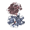
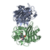
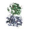
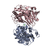
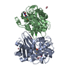
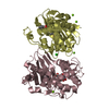
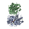
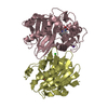
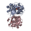
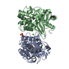
| Title | OXA-48 covalent complex with Avibactam inhibitor | |||||||||
 Components Components | (Beta-lactamase) x 2 | |||||||||
 Keywords Keywords | HYDROLASE / OXA-48 / Class D carbapenemase / Avibactam / inhibitor | |||||||||
| Function / homology |  Function and homology information Function and homology informationpenicillin binding / antibiotic catabolic process / cell wall organization / beta-lactamase activity / beta-lactamase / response to antibiotic / plasma membrane Similarity search - Function | |||||||||
| Biological species |  Klebsiella pneumoniae (bacteria) Klebsiella pneumoniae (bacteria) | |||||||||
| Method |  X-RAY DIFFRACTION / SYNCHROTRON / MOLECULAR REPLACEMENT / Resolution: 2.3 Å X-RAY DIFFRACTION / SYNCHROTRON / MOLECULAR REPLACEMENT / Resolution: 2.3 Å | |||||||||
 Authors Authors | Mangani, S. / Benvenuti, M. / Docquier, J.D. | |||||||||
 Citation Citation | Journal: Acs Chem.Biol. / Year: 2015 Title: Molecular Basis of Selective Inhibition and Slow Reversibility of Avibactam against Class D Carbapenemases: A Structure-Guided Study of OXA-24 and OXA-48. Authors: Lahiri, S.D. / Mangani, S. / Jahic, H. / Benvenuti, M. / Durand-Reville, T.F. / De Luca, F. / Ehmann, D.E. / Rossolini, G.M. / Alm, R.A. / Docquier, J.D. | |||||||||
| History |
|
- Structure visualization
| Structure viewer | Molecule: MolmilJmol/JSmol |
|---|
- Downloads & links
-Download
| PDBx/mmCIF format | 4wmc.cif.gz | 387.6 KB | Display | PDBx/mmCIF format |
|---|---|---|---|---|
| PDB format | pdb4wmc.ent.gz | 316.4 KB | Display | PDB format |
| PDBx/mmJSON format | 4wmc.json.gz | Tree view | PDBx/mmJSON format | |
| Others |  Other downloads Other downloads |
-Validation report
| Arichive directory | https://data.pdbj.org/pub/pdb/validation_reports/wm/4wmcftp://data.pdbj.org/pub/pdb/validation_reports/wm/4wmc | HTTPS FTP |
|---|
-Related structure data
| Related structure data | 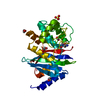 4wm9C  3hbrS S: Starting model for refinement C: citing same article ( |
|---|---|
| Similar structure data |


-Links
 PDBj
PDBj
- Assembly
| Deposited unit | 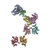
| ||||||||
|---|---|---|---|---|---|---|---|---|---|
| 1 | 
| ||||||||
| 2 | 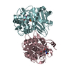
| ||||||||
| 3 | 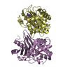
| ||||||||
| 4 | 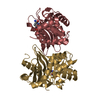
| ||||||||
| Unit cell |
|
-Components
| #1: Protein | Mass: 28054.768 Da / Num. of mol.: 6 / Fragment: UNP residues 24-265 Source method: isolated from a genetically manipulated source Source: (gene. exp.) Klebsiella pneumoniae (bacteria) / Gene: bla OXA-48, blaOXA-48, KPE71T_00045 / Production host: #2: Protein | Mass: 28097.770 Da / Num. of mol.: 2 / Fragment: UNP residues 24-265 Source method: isolated from a genetically manipulated source Source: (gene. exp.) Klebsiella pneumoniae (bacteria) / Gene: bla OXA-48, blaOXA-48, KPE71T_00045 / Production host: #3: Chemical | ChemComp-NXL / (   Mass: 267.260 Da / Num. of mol.: 8 / Source method: obtained synthetically / Formula: C7H13N3O6S / Comment: antibiotic, inhibitor*YM Mass: 267.260 Da / Num. of mol.: 8 / Source method: obtained synthetically / Formula: C7H13N3O6S / Comment: antibiotic, inhibitor*YM#4: Chemical |   Mass: 44.010 Da / Num. of mol.: 3 / Source method: obtained synthetically / Formula: CO2 Mass: 44.010 Da / Num. of mol.: 3 / Source method: obtained synthetically / Formula: CO2#5: Water | ChemComp-HOH / |  Mass: 18.015 Da / Num. of mol.: 256 / Source method: isolated from a natural source / Formula: H2O Mass: 18.015 Da / Num. of mol.: 256 / Source method: isolated from a natural source / Formula: H2O |
|---|
-Experimental details
-Experiment
| Experiment | Method: X-RAY DIFFRACTION / Number of used crystals: 1 |
|---|
- Sample preparation
| Crystal | Density Matthews: 2.56 Å3/Da / Density % sol: 51.87 % |
|---|---|
| Crystal grow | Temperature: 293 K / Method: vapor diffusion, sitting drop / pH: 7.5 Details: 0.1 M HEPES, 10% PEG8000, 5-8% 1-butanol, 10 mg/mL OXA-48, and 3 mg/mL avibactam |
-Data collection
| Diffraction | Mean temperature: 100 K |
|---|---|
| Diffraction source | Source: SYNCHROTRON / Site: ESRF  / Beamline: ID23-1 / Wavelength: 0.9834 Å / Beamline: ID23-1 / Wavelength: 0.9834 Å |
| Detector | Type: ADSC QUANTUM 315 / Detector: CCD / Date: Oct 25, 2008 |
| Radiation | Protocol: SINGLE WAVELENGTH / Monochromatic (M) / Laue (L): M / Scattering type: x-ray |
| Radiation wavelength | Wavelength: 0.9834 Å / Relative weight: 1 |
| Reflection | Resolution: 2.3→65.8 Å / Num. obs: 99733 / % possible obs: 93.4 % / Observed criterion σ(I): 2 / Redundancy: 3.1 % / Rmerge(I) obs: 0.068 / Net I/σ(I): 10.6 |
| Reflection shell | Resolution: 2.3→2.42 Å / Redundancy: 3 % / Rmerge(I) obs: 0.25 / Mean I/σ(I) obs: 5.9 / Num. unique all: 12691 / % possible all: 88 |
- Processing
| Software |
| ||||||||||||||||||||||||||||||||||||||||||||||||||||||||||||
|---|---|---|---|---|---|---|---|---|---|---|---|---|---|---|---|---|---|---|---|---|---|---|---|---|---|---|---|---|---|---|---|---|---|---|---|---|---|---|---|---|---|---|---|---|---|---|---|---|---|---|---|---|---|---|---|---|---|---|---|---|---|
| Refinement | Method to determine structure: MOLECULAR REPLACEMENT Starting model: 3HBR Resolution: 2.3→63.88 Å / Cor.coef. Fo:Fc: 0.915 / Cor.coef. Fo:Fc free: 0.862 / SU B: 8.172 / SU ML: 0.201 / Cross valid method: THROUGHOUT / σ(F): 0 / ESU R: 0.418 / ESU R Free: 0.281 / Stereochemistry target values: MAXIMUM LIKELIHOOD / Details: U VALUES : REFINED INDIVIDUALLY
| ||||||||||||||||||||||||||||||||||||||||||||||||||||||||||||
| Solvent computation | Ion probe radii: 0.8 Å / Shrinkage radii: 0.8 Å / VDW probe radii: 1.2 Å / Solvent model: MASK | ||||||||||||||||||||||||||||||||||||||||||||||||||||||||||||
| Displacement parameters | Biso max: 90 Å2 / Biso mean: 28.71 Å2 / Biso min: 2 Å2
| ||||||||||||||||||||||||||||||||||||||||||||||||||||||||||||
| Refinement step | Cycle: final / Resolution: 2.3→63.88 Å
| ||||||||||||||||||||||||||||||||||||||||||||||||||||||||||||
| Refine LS restraints |
| ||||||||||||||||||||||||||||||||||||||||||||||||||||||||||||
| LS refinement shell | Resolution: 2.3→2.36 Å / Total num. of bins used: 20
|
