- PDB-4wj1: Crystal structure of EspB from the ESX-1 type VII secretion system -
+
Open data
ID or keywords:
Loading...
-
Basic information
Entry
Database: PDB / ID: 4wj1
Title
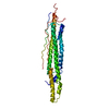
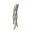
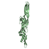
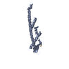
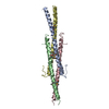
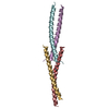
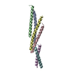
Crystal structure of EspB from the ESX-1 type VII secretion system
Components
Antigen MTB48, Mycobacterial protein
Keywords
PROTEIN TRANSPORT / Mycobacterial protein
Function / homology
ESX-1 secretion-associated protein EspB, PE domain / : / ESX-1 secreted protein B PE domain / ESX-1 secretion-associated protein EspB, PPE domain / identical protein binding / Antigen MTB48
Function and homology information
Biological species
Mycobacterium smegmatis (bacteria)
Method
X-RAY DIFFRACTION / SYNCHROTRON / Resolution: 2.415 Å
Journal: Structure / Year: 2015 Title: Structure of EspB from the ESX-1 type VII secretion system and insights into its export mechanism. Authors: Matthew Solomonson / Dheva Setiaputra / Karl A T Makepeace / Emilie Lameignere / Evgeniy V Petrotchenko / Deborah G Conrady / Julien R Bergeron / Marija Vuckovic / Frank DiMaio / Christoph H ...Authors: Matthew Solomonson / Dheva Setiaputra / Karl A T Makepeace / Emilie Lameignere / Evgeniy V Petrotchenko / Deborah G Conrady / Julien R Bergeron / Marija Vuckovic / Frank DiMaio / Christoph H Borchers / Calvin K Yip / Natalie C J Strynadka / Abstract: Mycobacterium tuberculosis (Mtb) uses the ESX-1 type VII secretion system to export virulence proteins across its lipid-rich cell wall, which helps permeabilize the host's macrophage phagosomal ...Mycobacterium tuberculosis (Mtb) uses the ESX-1 type VII secretion system to export virulence proteins across its lipid-rich cell wall, which helps permeabilize the host's macrophage phagosomal membrane, facilitating the escape and cell-to-cell spread of Mtb. ESX-1 membranolytic activity depends on a set of specialized secreted Esp proteins, the structure and specific roles of which are not currently understood. Here, we report the X-ray and electron microscopic structures of the ESX-1-secreted EspB. We demonstrate that EspB adopts a PE/PPE-like fold that mediates oligomerization with apparent heptameric symmetry, generating a barrel-shaped structure with a central pore that we propose contributes to the macrophage killing functions of EspB. Our structural data also reveal unexpected direct interactions between the EspB bipartite secretion signal sequence elements that form a unified aromatic surface. These findings provide insight into how specialized proteins encoded within the ESX-1 locus are targeted for secretion, and for the first time indicate an oligomerization-dependent role for Esp virulence factors.
In the structure databanks used in Yorodumi, some data are registered as the other names, "COVID-19 virus" and "2019-nCoV". Here are the details of the virus and the list of structure data.
Jan 31, 2019. EMDB accession codes are about to change! (news from PDBe EMDB page)
EMDB accession codes are about to change! (news from PDBe EMDB page)
The allocation of 4 digits for EMDB accession codes will soon come to an end. Whilst these codes will remain in use, new EMDB accession codes will include an additional digit and will expand incrementally as the available range of codes is exhausted. The current 4-digit format prefixed with “EMD-” (i.e. EMD-XXXX) will advance to a 5-digit format (i.e. EMD-XXXXX), and so on. It is currently estimated that the 4-digit codes will be depleted around Spring 2019, at which point the 5-digit format will come into force.
The EM Navigator/Yorodumi systems omit the EMD- prefix.
Related info.:Q: What is EMD? / ID/Accession-code notation in Yorodumi/EM Navigator
Yorodumi is a browser for structure data from EMDB, PDB, SASBDB, etc.
This page is also the successor to EM Navigator detail page, and also detail information page/front-end page for Omokage search.
The word "yorodu" (or yorozu) is an old Japanese word meaning "ten thousand". "mi" (miru) is to see.
Related info.:EMDB / PDB / SASBDB / Comparison of 3 databanks / Yorodumi Search / Aug 31, 2016. New EM Navigator & Yorodumi / Yorodumi Papers / Jmol/JSmol / Function and homology information / Changes in new EM Navigator and Yorodumi
 Movie
Movie Controller

 Yorodumi
 Open data
 Basic information
 Components
 Keywords
 Function and homology information
 Mycobacterium smegmatis (bacteria)
 X-RAY DIFFRACTION /
  Authors
 Citation

 Structure visualization
 Downloads & links
 Other downloads


 PDBj
PDBj Assembly
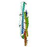
 Mass: 18.015 Da / Num. of mol.: 152 / Source method: isolated from a natural source / Formula: H2O
 Sample preparation
 Processing