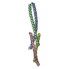
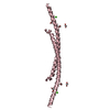
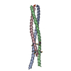
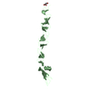
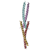
A: DNA binding ctp1 B: DNA binding ctp1 C: DNA binding ctp1 D: DNA binding ctp1 E: DNA binding ctp1 F: DNA binding ctp1 G: DNA binding ctp1 H: DNA binding ctp1 hetero molecules
DNAbindingctp1 / Double-strand break repair protein ctp1 / Meiotically up-regulated gene 38 protein / Nbs1- ...Double-strand break repair protein ctp1 / Meiotically up-regulated gene 38 protein / Nbs1-interacting protein 1 / Sporulation in the absence of SPO11 protein 2 homolog / SAE2
Mass: 6850.998 Da / Num. of mol.: 8 / Mutation: L51M Source method: isolated from a genetically manipulated source Source: (gene. exp.) Schizosaccharomyces pombe (fission yeast) Strain: 972 / ATCC 24843 / Gene: ctp1, mug38, nip1, slr9, SPCC338.08 / Plasmid: pMCSG9 / Production host: Escherichia coli (E. coli) References: UniProt: O74986, Hydrolases; Acting on ester bonds
Mass: 18.015 Da / Num. of mol.: 158 / Source method: isolated from a natural source / Formula: H2O
-
Experimental details
-
Experiment
Experiment
Method: X-RAY DIFFRACTION
-
Sample preparation
Crystal
Density Matthews: 3.01 Å3/Da / Density % sol: 59.2 %
Crystal grow
Temperature: 293 K / Method: vapor diffusion, hanging drop Details: Precipitant: 0.2M sodium acetate, 0.1M Tris pH 8.5 16% PEG 4000, with 0.4 uL of 0.1M barium chloride dihydrate additive. Cryoprotection in 25% ethylene glycol.
In the structure databanks used in Yorodumi, some data are registered as the other names, "COVID-19 virus" and "2019-nCoV". Here are the details of the virus and the list of structure data.
Jan 31, 2019. EMDB accession codes are about to change! (news from PDBe EMDB page)
EMDB accession codes are about to change! (news from PDBe EMDB page)
The allocation of 4 digits for EMDB accession codes will soon come to an end. Whilst these codes will remain in use, new EMDB accession codes will include an additional digit and will expand incrementally as the available range of codes is exhausted. The current 4-digit format prefixed with “EMD-” (i.e. EMD-XXXX) will advance to a 5-digit format (i.e. EMD-XXXXX), and so on. It is currently estimated that the 4-digit codes will be depleted around Spring 2019, at which point the 5-digit format will come into force.
The EM Navigator/Yorodumi systems omit the EMD- prefix.
Related info.:Q: What is EMD? / ID/Accession-code notation in Yorodumi/EM Navigator
Yorodumi is a browser for structure data from EMDB, PDB, SASBDB, etc.
This page is also the successor to EM Navigator detail page, and also detail information page/front-end page for Omokage search.
The word "yorodu" (or yorozu) is an old Japanese word meaning "ten thousand". "mi" (miru) is to see.
Related info.:EMDB / PDB / SASBDB / Comparison of 3 databanks / Yorodumi Search / Aug 31, 2016. New EM Navigator & Yorodumi / Yorodumi Papers / Jmol/JSmol / Function and homology information / Changes in new EM Navigator and Yorodumi
 Movie
Movie Controller


 Open data
 Basic information
 Components
 Keywords
 Function and homology information
 X-RAY DIFFRACTION /
  Authors
 United States, 1items
  Citation
 Structure visualization
 Downloads & links
 Other downloads
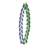


 PDBj
PDBj Assembly


 Mass: 62.068 Da / Num. of mol.: 13 / Source method: obtained synthetically / Formula: C2H6O2
 Mass: 18.015 Da / Num. of mol.: 158 / Source method: isolated from a natural source / Formula: H2O
 Sample preparation
 Processing