 Movie
Movie Controller

[English] 日本語
 Yorodumi
- PDB-4win: Crystal structure of the GATase domain from Plasmodium falciparum... -
+ Open data

- Basic information
| Entry | Database: PDB / ID: 4win | ||||||
|---|---|---|---|---|---|---|---|
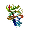
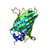
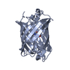
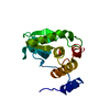
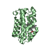
| Title | Crystal structure of the GATase domain from Plasmodium falciparum GMP synthetase | ||||||
 Components Components | GMP synthetase | ||||||
 Keywords Keywords | TRANSFERASE / GMP synthetase / Plasmodium falciparum / purine salvage pathway / glutamine amidotransferase | ||||||
| Function / homology |  Function and homology information Function and homology informationPurine ribonucleoside monophosphate biosynthesis / GMP synthase (glutamine-hydrolyzing) activity / GMP synthase activity / Azathioprine ADME / GMP synthase (glutamine-hydrolysing) / purine nucleotide biosynthetic process / GMP biosynthetic process / magnesium ion binding / ATP binding / cytosol Similarity search - Function | ||||||
| Biological species |  | ||||||
| Method |  X-RAY DIFFRACTION / SYNCHROTRON / MOLECULAR REPLACEMENT / Resolution: 2.6 Å X-RAY DIFFRACTION / SYNCHROTRON / MOLECULAR REPLACEMENT / Resolution: 2.6 Å | ||||||
 Authors Authors | Ballut, L. / Violot, S. / Haser, R. / Aghajari, N. | ||||||
 Citation Citation | Journal: Nat Commun / Year: 2015 Title: Active site coupling in Plasmodium falciparum GMP synthetase is triggered by domain rotation. Authors: Ballut, L. / Violot, S. / Shivakumaraswamy, S. / Thota, L.P. / Sathya, M. / Kunala, J. / Dijkstra, B.W. / Terreux, R. / Haser, R. / Balaram, H. / Aghajari, N. | ||||||
| History |
|
- Structure visualization
| Structure viewer | Molecule: MolmilJmol/JSmol |
|---|
- Downloads & links
-Download
| PDBx/mmCIF format | 4win.cif.gz | 104.1 KB | Display | PDBx/mmCIF format |
|---|---|---|---|---|
| PDB format | pdb4win.ent.gz | 78.6 KB | Display | PDB format |
| PDBx/mmJSON format | 4win.json.gz | Tree view | PDBx/mmJSON format | |
| Others |  Other downloads Other downloads |
-Validation report
| Arichive directory | https://data.pdbj.org/pub/pdb/validation_reports/wi/4winftp://data.pdbj.org/pub/pdb/validation_reports/wi/4win | HTTPS FTP |
|---|
-Related structure data
| Related structure data |  4wimC  4wioC  1gpmS C: citing same article ( S: Starting model for refinement |
|---|---|
| Similar structure data |


-Links
 PDBj
PDBj

- Assembly
| Deposited unit | 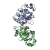
| ||||||||
|---|---|---|---|---|---|---|---|---|---|
| 1 | 
| ||||||||
| 2 | 
| ||||||||
| Unit cell |
|
-Components
| #1: Protein | Mass: 28829.703 Da / Num. of mol.: 2 Source method: isolated from a genetically manipulated source Source: (gene. exp.) Strain: isolate 3D7 / Gene: PF10_0123 / Plasmid: pET-21b(+) / Production host:  References: UniProt: Q8IJR9, GMP synthase (glutamine-hydrolysing) #2: Chemical |   Mass: 62.005 Da / Num. of mol.: 3 / Source method: obtained synthetically / Formula: NO3 Mass: 62.005 Da / Num. of mol.: 3 / Source method: obtained synthetically / Formula: NO3#3: Water | ChemComp-HOH / |  Mass: 18.015 Da / Num. of mol.: 93 / Source method: isolated from a natural source / Formula: H2O Mass: 18.015 Da / Num. of mol.: 93 / Source method: isolated from a natural source / Formula: H2O |
|---|
-Experimental details
-Experiment
| Experiment | Method: X-RAY DIFFRACTION |
|---|
- Sample preparation
| Crystal | Density Matthews: 2.09 Å3/Da / Density % sol: 41 % |
|---|---|
| Crystal grow | Temperature: 292 K / Method: vapor diffusion, sitting drop / pH: 7.4 / Details: 0.2 M Lithium Nitrate and 20% (w/v) PEG3350 |
-Data collection
| Diffraction | Mean temperature: 100 K |
|---|---|
| Diffraction source | Source: SYNCHROTRON / Site: ESRF  / Beamline: ID14-4 / Wavelength: 0.93928 Å / Beamline: ID14-4 / Wavelength: 0.93928 Å |
| Detector | Type: ADSC QUANTUM 315r / Detector: CCD / Date: Jul 10, 2011 |
| Radiation | Protocol: SINGLE WAVELENGTH / Monochromatic (M) / Laue (L): M / Scattering type: x-ray |
| Radiation wavelength | Wavelength: 0.93928 Å / Relative weight: 1 |
| Reflection | Resolution: 2.6→56 Å / Num. obs: 15347 / % possible obs: 99.6 % / Redundancy: 5.1 % / Net I/σ(I): 22.4 |
| Reflection shell | Resolution: 2.6→2.8 Å / Redundancy: 5 % / Rmerge(I) obs: 0.378 / Mean I/σ(I) obs: 4.67 / % possible all: 99.8 |
- Processing
| Software |
| ||||||||||||||||||||||||||||||||||||||||||
|---|---|---|---|---|---|---|---|---|---|---|---|---|---|---|---|---|---|---|---|---|---|---|---|---|---|---|---|---|---|---|---|---|---|---|---|---|---|---|---|---|---|---|---|
| Refinement | Method to determine structure: MOLECULAR REPLACEMENT Starting model: 1GPM Resolution: 2.6→38.972 Å / SU ML: 0.33 / Cross valid method: FREE R-VALUE / σ(F): 1.99 / Phase error: 23.51 / Stereochemistry target values: ML
| ||||||||||||||||||||||||||||||||||||||||||
| Solvent computation | Shrinkage radii: 0.9 Å / VDW probe radii: 1.11 Å / Solvent model: FLAT BULK SOLVENT MODEL | ||||||||||||||||||||||||||||||||||||||||||
| Refinement step | Cycle: LAST / Resolution: 2.6→38.972 Å
| ||||||||||||||||||||||||||||||||||||||||||
| Refine LS restraints |
| ||||||||||||||||||||||||||||||||||||||||||
| LS refinement shell |
|
