 Movie
Movie Controller

[English] 日本語
 Yorodumi
- PDB-5uxg: Protein 84 with aldehyde deformylating oxygenase activity from Su... -
+ Open data

- Basic information
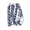
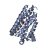
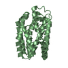
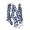
| Entry | Database: PDB / ID: 5uxg | ||||||
|---|---|---|---|---|---|---|---|
| Title | Protein 84 with aldehyde deformylating oxygenase activity from Sulfolobus tokodaii (monoclinic) | ||||||
 Components Components | aldehyde deformylating oxygenase | ||||||
 Keywords Keywords | LYASE / decarbonylase / iron / ferritin | ||||||
| Function / homology | Ribonucleotide reductase-like / Ferritin-like superfamily / oxidoreductase activity / metal ion binding / PHOSPHATE ION / Uncharacterized protein Function and homology information Function and homology information | ||||||
| Biological species |   Sulfolobus tokodaii (archaea) Sulfolobus tokodaii (archaea) | ||||||
| Method |  X-RAY DIFFRACTION / SYNCHROTRON / MOLECULAR REPLACEMENT / Resolution: 1.72 Å X-RAY DIFFRACTION / SYNCHROTRON / MOLECULAR REPLACEMENT / Resolution: 1.72 Å | ||||||
 Authors Authors | Wilson, D.K. / Mak, W.S. / Siegel, J.B. | ||||||
 Citation Citation | Journal: To Be Published Title: Protein 84 with aldehyde deformylating oxygenase activity from Sulfolobus tokodaii (monoclinic) Authors: Wilson, D.K. / Mak, W.S. / Siegel, J.B. | ||||||
| History |
|
- Structure visualization
| Structure viewer | Molecule: MolmilJmol/JSmol |
|---|
- Downloads & links
-Download
| PDBx/mmCIF format | 5uxg.cif.gz | 102.4 KB | Display | PDBx/mmCIF format |
|---|---|---|---|---|
| PDB format | pdb5uxg.ent.gz | 78.9 KB | Display | PDB format |
| PDBx/mmJSON format | 5uxg.json.gz | Tree view | PDBx/mmJSON format | |
| Others |  Other downloads Other downloads |
-Validation report
| Arichive directory | https://data.pdbj.org/pub/pdb/validation_reports/ux/5uxgftp://data.pdbj.org/pub/pdb/validation_reports/ux/5uxg | HTTPS FTP |
|---|
-Related structure data
| Related structure data | |
|---|---|
| Similar structure data |


-Links
 PDBj
PDBj

- Assembly
| Deposited unit | 
| ||||||||
|---|---|---|---|---|---|---|---|---|---|
| 1 | 
| ||||||||
| 2 | 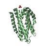
| ||||||||
| Unit cell |
|
-Components
| #1: Protein | Mass: 25917.844 Da / Num. of mol.: 2 Source method: isolated from a genetically manipulated source Source: (gene. exp.) Sulfolobus tokodaii (strain DSM 16993 / JCM 10545 / NBRC 100140 / 7) (archaea)Strain: DSM 16993 / JCM 10545 / NBRC 100140 / 7 / Gene: ST1620, STK_16200 / Production host:  #2: Chemical |   Mass: 94.971 Da / Num. of mol.: 3 / Source method: obtained synthetically / Formula: PO4 Mass: 94.971 Da / Num. of mol.: 3 / Source method: obtained synthetically / Formula: PO4#3: Water | ChemComp-HOH / |  Mass: 18.015 Da / Num. of mol.: 212 / Source method: isolated from a natural source / Formula: H2O Mass: 18.015 Da / Num. of mol.: 212 / Source method: isolated from a natural source / Formula: H2O |
|---|
-Experimental details
-Experiment
| Experiment | Method: X-RAY DIFFRACTION / Number of used crystals: 1 |
|---|
- Sample preparation
| Crystal | Density Matthews: 2.54 Å3/Da / Density % sol: 51.52 % |
|---|---|
| Crystal grow | Temperature: 298 K / Method: vapor diffusion, hanging drop / Details: Potassium phosphate, PEG 8000 |
-Data collection
| Diffraction | Mean temperature: 100 K |
|---|---|
| Diffraction source | Source: SYNCHROTRON / Site: SSRL  / Beamline: BL9-2 / Wavelength: 0.97946 Å / Beamline: BL9-2 / Wavelength: 0.97946 Å |
| Detector | Type: DECTRIS PILATUS3 6M / Detector: PIXEL / Date: May 27, 2016 |
| Radiation | Protocol: SINGLE WAVELENGTH / Monochromatic (M) / Laue (L): M / Scattering type: x-ray |
| Radiation wavelength | Wavelength: 0.97946 Å / Relative weight: 1 |
| Reflection | Resolution: 1.72→63.61 Å / Num. obs: 55207 / % possible obs: 96.5 % / Redundancy: 6.7 % / Rmerge(I) obs: 0.05 / Net I/σ(I): 20.2 |
| Reflection shell | Resolution: 1.72→1.75 Å / Rmerge(I) obs: 0.251 / Mean I/σ(I) obs: 3.7 / % possible all: 57 |
- Processing
| Software |
| ||||||||||||||||||||||||||||||||||||||||||||||||||||||||||||||||||||||||||||||||||||||||||||||||||||||||||||||||||||||||||||||||||||||||||||||||||||||||||||||||||||||||||||||||||||||
|---|---|---|---|---|---|---|---|---|---|---|---|---|---|---|---|---|---|---|---|---|---|---|---|---|---|---|---|---|---|---|---|---|---|---|---|---|---|---|---|---|---|---|---|---|---|---|---|---|---|---|---|---|---|---|---|---|---|---|---|---|---|---|---|---|---|---|---|---|---|---|---|---|---|---|---|---|---|---|---|---|---|---|---|---|---|---|---|---|---|---|---|---|---|---|---|---|---|---|---|---|---|---|---|---|---|---|---|---|---|---|---|---|---|---|---|---|---|---|---|---|---|---|---|---|---|---|---|---|---|---|---|---|---|---|---|---|---|---|---|---|---|---|---|---|---|---|---|---|---|---|---|---|---|---|---|---|---|---|---|---|---|---|---|---|---|---|---|---|---|---|---|---|---|---|---|---|---|---|---|---|---|---|---|
| Refinement | Method to determine structure: MOLECULAR REPLACEMENT Starting model: theoretical Resolution: 1.72→63.61 Å / Cor.coef. Fo:Fc: 0.945 / Cor.coef. Fo:Fc free: 0.93 / SU B: 2.494 / SU ML: 0.082 / Cross valid method: THROUGHOUT / ESU R: 0.11 / ESU R Free: 0.106 / Details: HYDROGENS HAVE BEEN ADDED IN THE RIDING POSITIONS
| ||||||||||||||||||||||||||||||||||||||||||||||||||||||||||||||||||||||||||||||||||||||||||||||||||||||||||||||||||||||||||||||||||||||||||||||||||||||||||||||||||||||||||||||||||||||
| Solvent computation | Ion probe radii: 0.8 Å / Shrinkage radii: 0.8 Å / VDW probe radii: 1.2 Å | ||||||||||||||||||||||||||||||||||||||||||||||||||||||||||||||||||||||||||||||||||||||||||||||||||||||||||||||||||||||||||||||||||||||||||||||||||||||||||||||||||||||||||||||||||||||
| Displacement parameters | Biso mean: 22.536 Å2
| ||||||||||||||||||||||||||||||||||||||||||||||||||||||||||||||||||||||||||||||||||||||||||||||||||||||||||||||||||||||||||||||||||||||||||||||||||||||||||||||||||||||||||||||||||||||
| Refinement step | Cycle: 1 / Resolution: 1.72→63.61 Å
| ||||||||||||||||||||||||||||||||||||||||||||||||||||||||||||||||||||||||||||||||||||||||||||||||||||||||||||||||||||||||||||||||||||||||||||||||||||||||||||||||||||||||||||||||||||||
| Refine LS restraints |
|