 Movie
Movie Controller

[English] 日本語
 Yorodumi
- PDB-6n14: Phosphoserine BlaC, Class A serine beta-lactamase from Mycobacter... -
+ Open data

- Basic information
| Entry | Database: PDB / ID: 6n14 | ||||||
|---|---|---|---|---|---|---|---|
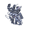
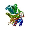
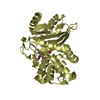
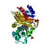
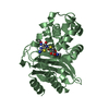
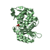
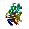
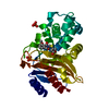
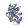
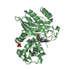
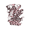
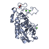
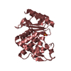
| Title | Phosphoserine BlaC, Class A serine beta-lactamase from Mycobacterium tuberculosis | ||||||
 Components Components | Beta-lactamase | ||||||
 Keywords Keywords | HYDROLASE / phosphoserine / beta-lactamase / Mycobacterium tuberculosis / inhibitor | ||||||
| Function / homology |  Function and homology information Function and homology informationbeta-lactam antibiotic catabolic process / beta-lactamase activity / beta-lactamase / periplasmic space / response to antibiotic / extracellular region / plasma membrane Similarity search - Function | ||||||
| Biological species |   Mycobacterium tuberculosis (bacteria) Mycobacterium tuberculosis (bacteria) | ||||||
| Method |  X-RAY DIFFRACTION / SYNCHROTRON / MOLECULAR REPLACEMENT / Resolution: 1.52169460473 Å X-RAY DIFFRACTION / SYNCHROTRON / MOLECULAR REPLACEMENT / Resolution: 1.52169460473 Å | ||||||
 Authors Authors | Moural, T.W. / White, D.S. / Choy, C.J. / Kang, C. / Berkman, C.E. | ||||||
 Citation Citation | Journal: Int J Mol Sci / Year: 2019 Title: Crystal Structure of Phosphoserine BlaC fromMycobacterium tuberculosisInactivated by Bis(Benzoyl) Phosphate. Authors: Moural, T.W. / White, D.S. / Choy, C.J. / Kang, C. / Berkman, C.E. | ||||||
| History |
|
- Structure visualization
| Structure viewer | Molecule: MolmilJmol/JSmol |
|---|
- Downloads & links
-Download
| PDBx/mmCIF format | 6n14.cif.gz | 139.3 KB | Display | PDBx/mmCIF format |
|---|---|---|---|---|
| PDB format | pdb6n14.ent.gz | 87.5 KB | Display | PDB format |
| PDBx/mmJSON format | 6n14.json.gz | Tree view | PDBx/mmJSON format | |
| Others |  Other downloads Other downloads |
-Validation report
| Arichive directory | https://data.pdbj.org/pub/pdb/validation_reports/n1/6n14ftp://data.pdbj.org/pub/pdb/validation_reports/n1/6n14 | HTTPS FTP |
|---|
-Related structure data
| Related structure data | 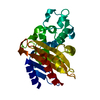 2gdnS S: Starting model for refinement |
|---|---|
| Similar structure data |


-Links
 PDBj
PDBj


- Assembly
| Deposited unit | 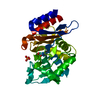
| ||||||||||||
|---|---|---|---|---|---|---|---|---|---|---|---|---|---|
| 1 |
| ||||||||||||
| Unit cell |
|
-Components
| #1: Protein | Mass: 32699.791 Da / Num. of mol.: 1 Source method: isolated from a genetically manipulated source Source: (gene. exp.) Mycobacterium tuberculosis (bacteria) / Gene: blaC, ERS027646_02769 / Plasmid: pET28 / Production host: References: UniProt: A0A0T9EA39, UniProt: P9WKD3*PLUS, beta-lactamase | ||||
|---|---|---|---|---|---|
| #2: Chemical |   Mass: 94.971 Da / Num. of mol.: 2 / Source method: obtained synthetically / Formula: PO4 Mass: 94.971 Da / Num. of mol.: 2 / Source method: obtained synthetically / Formula: PO4#3: Water | ChemComp-HOH / |  Mass: 18.015 Da / Num. of mol.: 329 / Source method: isolated from a natural source / Formula: H2O Mass: 18.015 Da / Num. of mol.: 329 / Source method: isolated from a natural source / Formula: H2OHas protein modification | Y | |
-Experimental details
-Experiment
| Experiment | Method: X-RAY DIFFRACTION / Number of used crystals: 1 |
|---|
- Sample preparation
| Crystal | Density Matthews: 1.96 Å3/Da / Density % sol: 37.12 % |
|---|---|
| Crystal grow | Temperature: 277.15 K / Method: vapor diffusion, hanging drop / pH: 7.4 Details: 0.1 M HEPES pH 7.4, 2.25 M ammonium phosphate monobasic, protein concertation 15 mg/mL |
-Data collection
| Diffraction | Mean temperature: 100 K / Serial crystal experiment: N |
|---|---|
| Diffraction source | Source: SYNCHROTRON / Site: ALS  / Beamline: 8.2.2 / Wavelength: 1 Å / Beamline: 8.2.2 / Wavelength: 1 Å |
| Detector | Type: ADSC QUANTUM 315r / Detector: CCD / Date: Nov 17, 2017 |
| Radiation | Monochromator: Double-crystal Si(111) / Protocol: SINGLE WAVELENGTH / Monochromatic (M) / Laue (L): M / Scattering type: x-ray |
| Radiation wavelength | Wavelength: 1 Å / Relative weight: 1 |
| Reflection | Resolution: 1.52→50 Å / Num. obs: 40045 / % possible obs: 99.9 % / Redundancy: 6.8 % / CC1/2: 0.992 / Rmerge(I) obs: 0.099 / Rpim(I) all: 0.04 / Rrim(I) all: 0.107 / Rsym value: 0.099 / Net I/σ(I): 17.586 |
| Reflection shell | Resolution: 1.52→1.56 Å / Mean I/σ(I) obs: 1.92 / CC1/2: 0.623 / Rpim(I) all: 0.403 / Rrim(I) all: 0.914 / % possible all: 98.9 |
- Processing
| Software |
| |||||||||||||||||||||||||||||||||||||||||||||||||||||||||||||||||||||||||||||||||||||||||||||||||||||||||
|---|---|---|---|---|---|---|---|---|---|---|---|---|---|---|---|---|---|---|---|---|---|---|---|---|---|---|---|---|---|---|---|---|---|---|---|---|---|---|---|---|---|---|---|---|---|---|---|---|---|---|---|---|---|---|---|---|---|---|---|---|---|---|---|---|---|---|---|---|---|---|---|---|---|---|---|---|---|---|---|---|---|---|---|---|---|---|---|---|---|---|---|---|---|---|---|---|---|---|---|---|---|---|---|---|---|---|
| Refinement | Method to determine structure: MOLECULAR REPLACEMENT Starting model: 2gdn Resolution: 1.52169460473→28.0973861897 Å / SU ML: 0.117484022888 / Cross valid method: THROUGHOUT / σ(F): 1.34370318253 / Phase error: 15.6852788604
| |||||||||||||||||||||||||||||||||||||||||||||||||||||||||||||||||||||||||||||||||||||||||||||||||||||||||
| Solvent computation | Shrinkage radii: 0.9 Å / VDW probe radii: 1.11 Å | |||||||||||||||||||||||||||||||||||||||||||||||||||||||||||||||||||||||||||||||||||||||||||||||||||||||||
| Displacement parameters | Biso mean: 16.7185917212 Å2 | |||||||||||||||||||||||||||||||||||||||||||||||||||||||||||||||||||||||||||||||||||||||||||||||||||||||||
| Refinement step | Cycle: LAST / Resolution: 1.52169460473→28.0973861897 Å
| |||||||||||||||||||||||||||||||||||||||||||||||||||||||||||||||||||||||||||||||||||||||||||||||||||||||||
| Refine LS restraints |
| |||||||||||||||||||||||||||||||||||||||||||||||||||||||||||||||||||||||||||||||||||||||||||||||||||||||||
| LS refinement shell |
|
