 Movie
Movie Controller

[English] 日本語
 Yorodumi
- PDB-4w2q: Anti-Marburgvirus Nucleoprotein Single Domain Antibody C Complexe... -
+ Open data

- Basic information
| Entry | Database: PDB / ID: 4w2q | ||||||
|---|---|---|---|---|---|---|---|
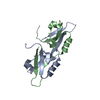
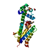
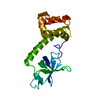
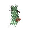
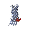
| Title | Anti-Marburgvirus Nucleoprotein Single Domain Antibody C Complexed with Nucleoprotein C-terminal domain | ||||||
 Components Components |
| ||||||
 Keywords Keywords | IMMUNE SYSTEM | ||||||
| Function / homology |  Function and homology information Function and homology informationviral RNA genome packaging / helical viral capsid / viral budding via host ESCRT complex / viral nucleocapsid / host cell cytoplasm / ribonucleoprotein complex / RNA binding Similarity search - Function | ||||||
| Biological species |   Lake Victoria marburgvirus Lake Victoria marburgvirus | ||||||
| Method |  X-RAY DIFFRACTION / MOLECULAR REPLACEMENT / Resolution: 2.7 Å X-RAY DIFFRACTION / MOLECULAR REPLACEMENT / Resolution: 2.7 Å | ||||||
 Authors Authors | Taylor, A.B. / Garza, J.A. | ||||||
 Citation Citation | Journal: Front Immunol / Year: 2017 Title: Unveiling a Drift Resistant Cryptotope withinMarburgvirusNucleoprotein Recognized by Llama Single-Domain Antibodies. Authors: Garza, J.A. / Taylor, A.B. / Sherwood, L.J. / Hart, P.J. / Hayhurst, A. | ||||||
| History |
|
- Structure visualization
| Structure viewer | Molecule: MolmilJmol/JSmol |
|---|
- Downloads & links
-Download
| PDBx/mmCIF format | 4w2q.cif.gz | 153.9 KB | Display | PDBx/mmCIF format |
|---|---|---|---|---|
| PDB format | pdb4w2q.ent.gz | 121.6 KB | Display | PDB format |
| PDBx/mmJSON format | 4w2q.json.gz | Tree view | PDBx/mmJSON format | |
| Others |  Other downloads Other downloads |
-Validation report
| Arichive directory | https://data.pdbj.org/pub/pdb/validation_reports/w2/4w2qftp://data.pdbj.org/pub/pdb/validation_reports/w2/4w2q | HTTPS FTP |
|---|
-Related structure data
| Related structure data |  4w2oC  4w2pC  6apoC 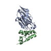 6appSC  6apqC S: Starting model for refinement C: citing same article ( |
|---|---|
| Similar structure data |


-Links
 PDBj
PDBj


- Assembly
| Deposited unit | 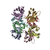
| ||||||||
|---|---|---|---|---|---|---|---|---|---|
| 1 | 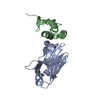
| ||||||||
| 2 | 
| ||||||||
| 3 | 
| ||||||||
| 4 | 
| ||||||||
| Unit cell |
|
-Components
| #1: Antibody | Mass: 12981.421 Da / Num. of mol.: 4 Source method: isolated from a genetically manipulated source Source: (gene. exp.) Description: Semi-synthetic single pot library Nomad 1 based upon Lama glama Plasmid: PECAN219 / Production host:  #2: Protein | Mass: 8768.806 Da / Num. of mol.: 4 / Fragment: C-terminal domain residues 632-695 Source method: isolated from a genetically manipulated source Source: (gene. exp.) Lake Victoria marburgvirus / Strain: Musoke-80 / Gene: NP / Plasmid: pE-NP632 / Production host: #3: Water | ChemComp-HOH / |  Mass: 18.015 Da / Num. of mol.: 104 / Source method: isolated from a natural source / Formula: H2O Mass: 18.015 Da / Num. of mol.: 104 / Source method: isolated from a natural source / Formula: H2OHas protein modification | Y | |
|---|
-Experimental details
-Experiment
| Experiment | Method: X-RAY DIFFRACTION / Number of used crystals: 1 |
|---|
- Sample preparation
| Crystal | Density Matthews: 2.24 Å3/Da / Density % sol: 45.01 % |
|---|---|
| Crystal grow | Temperature: 277 K / Method: vapor diffusion, sitting drop Details: 20% polyethylene glycol 6000, 0.2M magnesium chloride, 0.1M 1,2,3-hexanetriol, 0.1 M sodium acetate pH 5 |
-Data collection
| Diffraction | Mean temperature: 100 K |
|---|---|
| Diffraction source | Source: ROTATING ANODE / Type: RIGAKU MICROMAX-007 HF / Wavelength: 1.54178 Å |
| Detector | Type: RIGAKU RAXIS HTC / Detector: IMAGE PLATE / Date: Jun 1, 2016 |
| Radiation | Protocol: SINGLE WAVELENGTH / Monochromatic (M) / Laue (L): M / Scattering type: x-ray |
| Radiation wavelength | Wavelength: 1.54178 Å / Relative weight: 1 |
| Reflection | Resolution: 2.7→46.41 Å / Num. obs: 20770 / % possible obs: 99 % / Redundancy: 3.7 % / Biso Wilson estimate: 35.8 Å2 / Rpim(I) all: 0.092 / Rsym value: 0.154 / Net I/σ(I): 8.2 |
| Reflection shell | Resolution: 2.7→2.85 Å / Redundancy: 3.8 % / Mean I/σ(I) obs: 1.9 / Num. unique obs: 3013 / Rpim(I) all: 0.398 / Rsym value: 0.673 / % possible all: 98.3 |
- Processing
| Software |
| |||||||||||||||||||||||||||||||||||||||||||||||||||||||||||||||||||||||||||||||||||||||||||||||||||||||||
|---|---|---|---|---|---|---|---|---|---|---|---|---|---|---|---|---|---|---|---|---|---|---|---|---|---|---|---|---|---|---|---|---|---|---|---|---|---|---|---|---|---|---|---|---|---|---|---|---|---|---|---|---|---|---|---|---|---|---|---|---|---|---|---|---|---|---|---|---|---|---|---|---|---|---|---|---|---|---|---|---|---|---|---|---|---|---|---|---|---|---|---|---|---|---|---|---|---|---|---|---|---|---|---|---|---|---|
| Refinement | Method to determine structure: MOLECULAR REPLACEMENT Starting model: 6APP Resolution: 2.7→41.68 Å / SU ML: 0.41 / Cross valid method: THROUGHOUT / σ(F): 1.37 / Phase error: 28.88
| |||||||||||||||||||||||||||||||||||||||||||||||||||||||||||||||||||||||||||||||||||||||||||||||||||||||||
| Solvent computation | Shrinkage radii: 0.9 Å / VDW probe radii: 1 Å | |||||||||||||||||||||||||||||||||||||||||||||||||||||||||||||||||||||||||||||||||||||||||||||||||||||||||
| Displacement parameters | Biso mean: 38.7 Å2 | |||||||||||||||||||||||||||||||||||||||||||||||||||||||||||||||||||||||||||||||||||||||||||||||||||||||||
| Refinement step | Cycle: LAST / Resolution: 2.7→41.68 Å
| |||||||||||||||||||||||||||||||||||||||||||||||||||||||||||||||||||||||||||||||||||||||||||||||||||||||||
| Refine LS restraints |
| |||||||||||||||||||||||||||||||||||||||||||||||||||||||||||||||||||||||||||||||||||||||||||||||||||||||||
| LS refinement shell |
|
